Electron microscope chamber
1344×768 · AVIF · CC BY 4.0
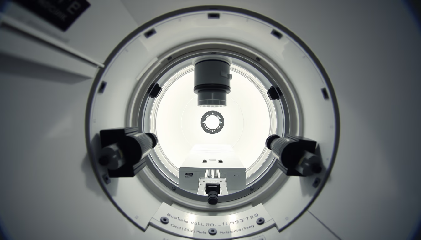
The chamber of an electron microscope is the vacuum-sealed compartment where the electron beam interacts with the sample, enabling magnifications of up to 2 million times.
About this subject
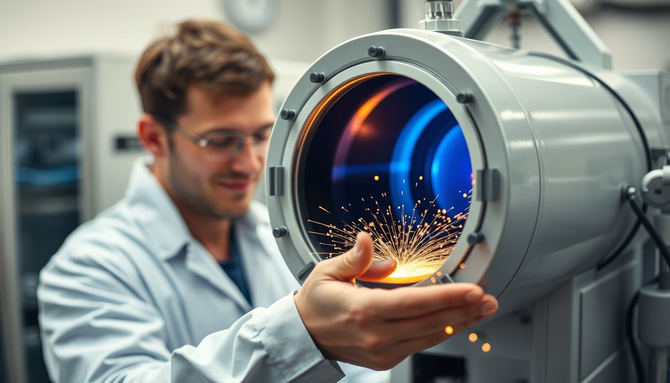
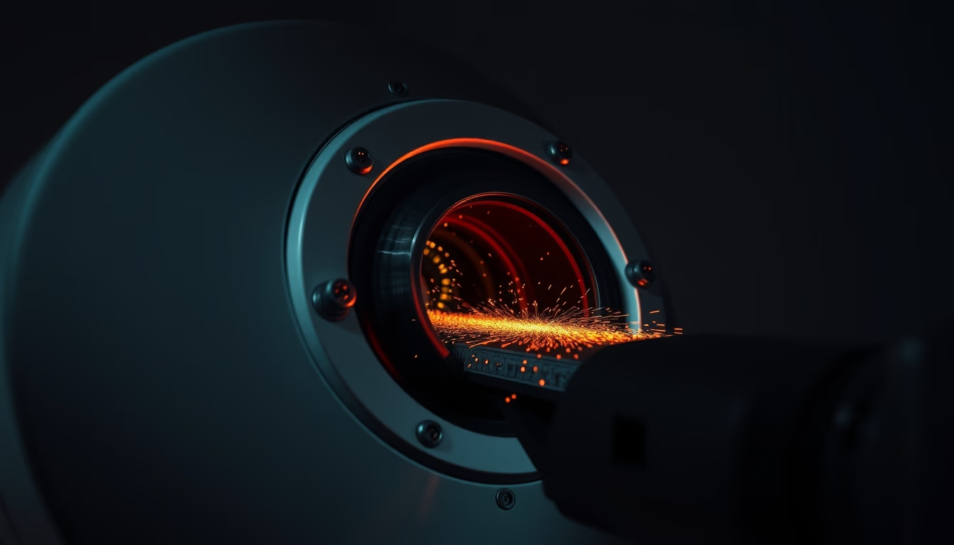
The chamber of an electron microscope is the central component, designed to maintain ultra-high vacuum conditions (in the range of 10⁻⁴ to 10⁻⁷ Pa). This vacuum is essential to prevent air molecules from scattering or deflecting the electron beam, ensuring high resolution. Inside the chamber, electromagnetic lenses and deflection coils focus and direct the beam onto the sample, while detectors capture secondary, backscattered, or transmitted electrons to form the image.
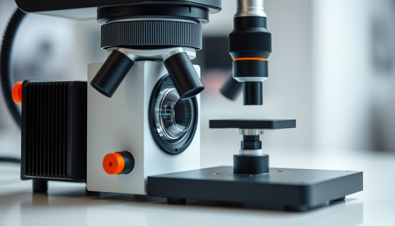
There are two main types of electron microscopes: scanning electron microscope (SEM) and transmission electron microscope (TEM). In SEM, the beam scans the sample surface and generates three-dimensional images with great depth of field, ideal for observing topography and composition. In TEM, the beam passes through an ultra-thin sample and reveals internal details at the atomic scale, such as crystalline defects and material structures.
Modern chambers are equipped with sample manipulation systems, access ports, and vacuum pumps (turbomolecular and ion pumps). To minimize vibrations, the chamber is rigidly mounted and may be isolated by air suspensions or springs. Additionally, internal coatings of materials like stainless steel or titanium reduce contamination. Regular cleaning and maintenance are critical to preserve the vacuum and avoid artifacts in images.
The importance of the chamber goes beyond simple containment: it enables advanced techniques such as energy-dispersive spectroscopy (EDS), electron backscatter diffraction (EBSD), and cryomicroscopy. In biology, cryogenic chambers maintain frozen hydrated samples, allowing the study of cellular structures in their native state. In materials science, the chamber enables failure analysis, coating inspection, and nanoparticle characterization with sub-nanometer resolution.
Frequently Asked Questions
Why does the electron microscope chamber need a vacuum?
The vacuum is necessary to prevent air molecules from interacting with the electron beam, which would cause scattering and loss of resolution. It also protects the filament from oxidation and contamination.
What is the difference between the chamber of an SEM and a TEM?
In SEM, the chamber houses detectors and allows scanning of the sample surface. In TEM, the chamber is longer to accommodate the transmitted beam path, and the sample must be ultra-thin for electrons to pass through.
How is the chamber maintained to avoid contamination?
The chamber is periodically cleaned with appropriate solvents and specialized equipment. Vacuum pumps run continuously, and seals are regularly inspected. Samples are introduced through auxiliary load chambers to minimize contamination.
Direct URL
https://pub-c7d6a6ea828543ac903a74a341ccb2e1.r2.dev/imagens/electron-microscope-chamber-fish-eye-wide-angle-fresh17.avifHow to credit
Include a visible link back to UtilizAí. Copy one of the snippets below:
<a href="https://xn--utiliza-eza.com/en/midia/imagens/electron-microscope-chamber-fish-eye-wide-angle-fresh17">Electron microscope chamber</a> by <a href="https://xn--utiliza-eza.com">UtilizAí</a>, licensed under <a href="https://creativecommons.org/licenses/by/4.0/">CC BY 4.0</a>.
[Electron microscope chamber](https://xn--utiliza-eza.com/en/midia/imagens/electron-microscope-chamber-fish-eye-wide-angle-fresh17) by [UtilizAí](https://xn--utiliza-eza.com), CC BY 4.0
License: CC-BY-4.0