Electron microscope chamber
1344×768 · AVIF · CC BY 4.0
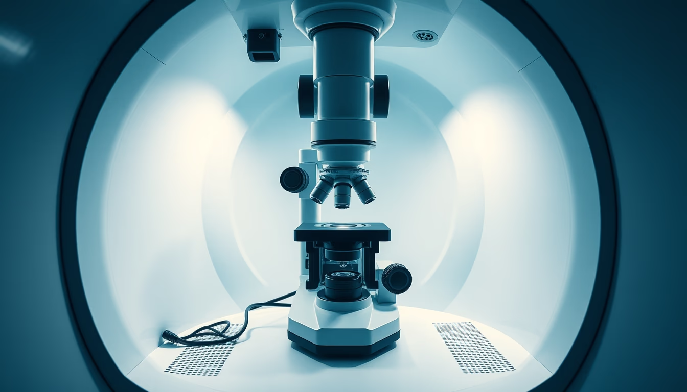
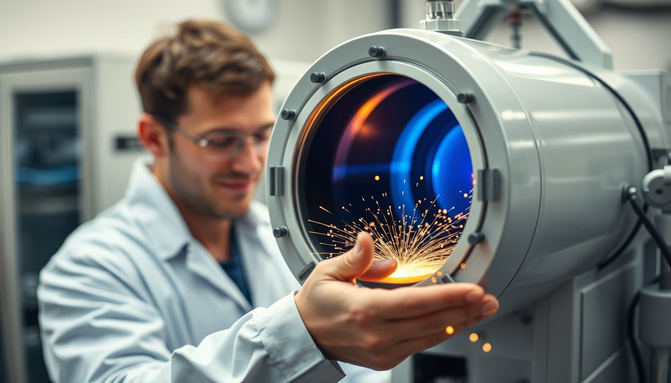
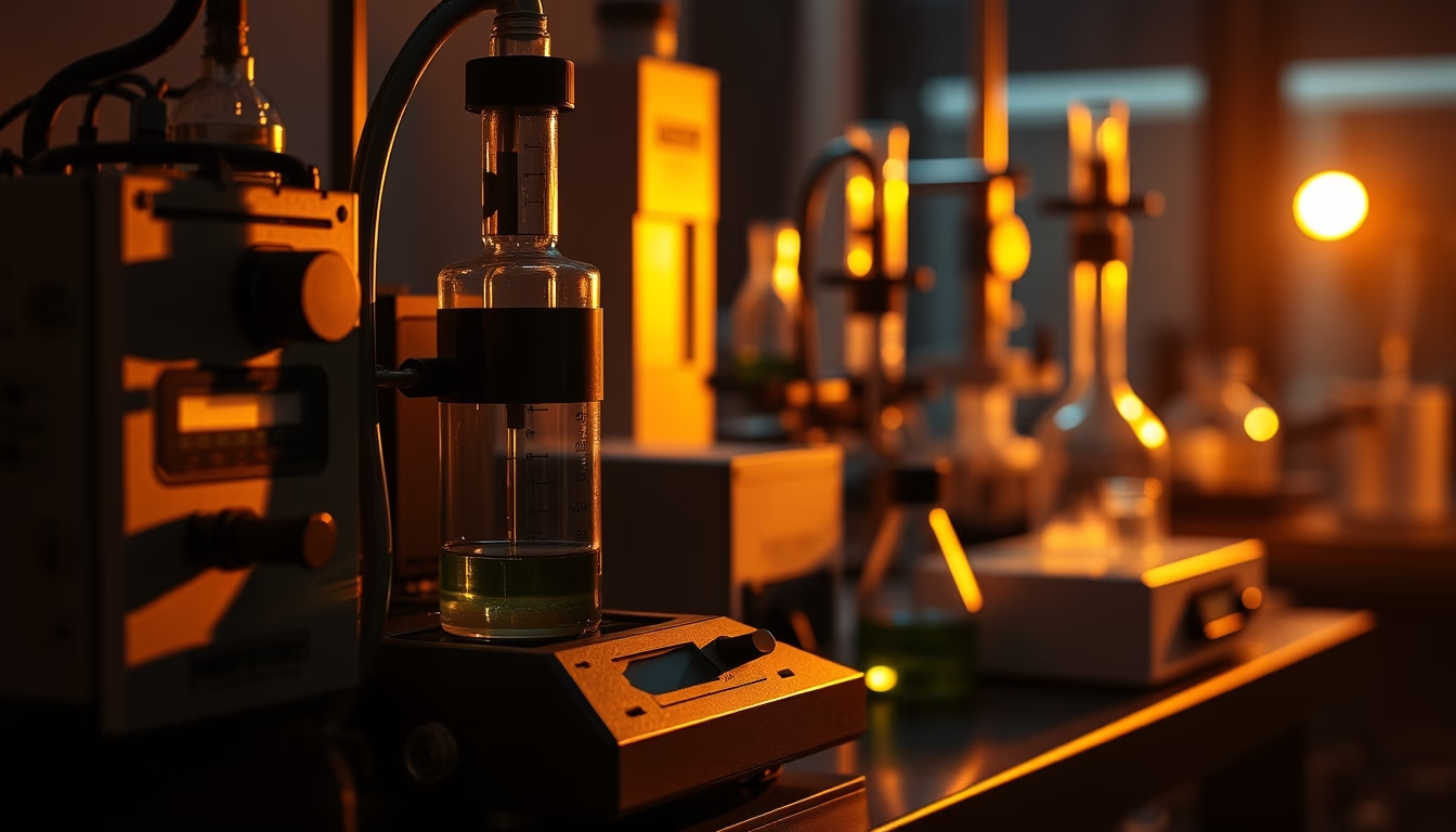
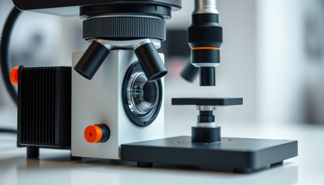
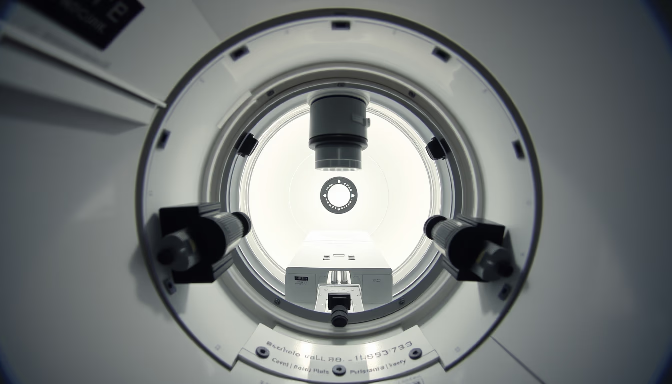
Electron microscope chamber, a critical component that maintains vacuum for electron beam analysis of specimens.
About this subject
The electron microscope chamber is the sealed compartment where the specimen is placed during analysis. It operates under high vacuum, typically between 10⁻⁴ and 10⁻⁷ Pa. This vacuum is essential to prevent air molecules from scattering or deflecting the electron beam, ensuring high resolution and contrast. The chamber is usually made of stainless steel or special alloys, with multiple windows and ports for detectors, manipulators, and cryogenic systems.
Early electron microscopes, developed by Ernst Ruska and Max Knoll in the 1930s, used rudimentary chambers with oil diffusion pumps. Modern chambers incorporate turbomolecular and ion pumps, reaching ultra-high vacuum in minutes. Fast-loading systems, such as airlocks, allow sample exchange without compromising the column vacuum.
The chamber houses the specimen stage, which can be heated, cooled, or tilted for multi-condition analyses. Detectors for secondary electrons, backscattered electrons, and X-rays are mounted on standardized flanges. In transmission electron microscopes (TEM), the chamber contains objective lenses and recording systems on film or digital cameras.
Chamber maintenance requires rigorous cleaning to avoid contamination, which degrades vacuum and image quality. Techniques like bake-out (heating for hours) remove adsorbed gases from walls. The chamber is also designed to minimize vibrations and parasitic electromagnetic fields, factors that limit the final resolution of the instrument.
Frequently Asked Questions
Why is vacuum necessary in the electron microscope chamber?
Vacuum prevents air molecules from interacting with the electron beam, causing scattering and loss of resolution. It also protects the filament and prevents electrical discharges.
What is the typical pressure inside the chamber?
Ranges from 10⁻⁴ Pa for scanning electron microscopes (SEM) to 10⁻⁷ Pa for high-resolution transmission electron microscopes (HRTEM).
How are samples inserted without breaking vacuum?
An airlock system is used: the sample is placed in an intermediate chamber, evacuated, then transferred to the main chamber without exposing the vacuum to the atmosphere.
Direct URL
https://pub-c7d6a6ea828543ac903a74a341ccb2e1.r2.dev/imagens/electron-microscope-chamber-minimalist-composition-fresh5.avifHow to credit
Include a visible link back to UtilizAí. Copy one of the snippets below:
<a href="https://xn--utiliza-eza.com/en/midia/imagens/electron-microscope-chamber-minimalist-composition-fresh5">Electron microscope chamber</a> by <a href="https://xn--utiliza-eza.com">UtilizAí</a>, licensed under <a href="https://creativecommons.org/licenses/by/4.0/">CC BY 4.0</a>.
[Electron microscope chamber](https://xn--utiliza-eza.com/en/midia/imagens/electron-microscope-chamber-minimalist-composition-fresh5) by [UtilizAí](https://xn--utiliza-eza.com), CC BY 4.0
License: CC-BY-4.0