Electron microscope chamber
1344×768 · AVIF · CC BY 4.0
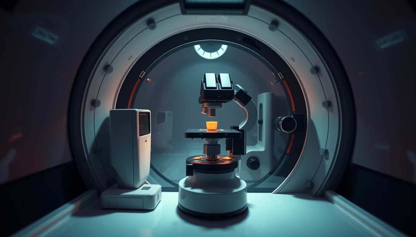
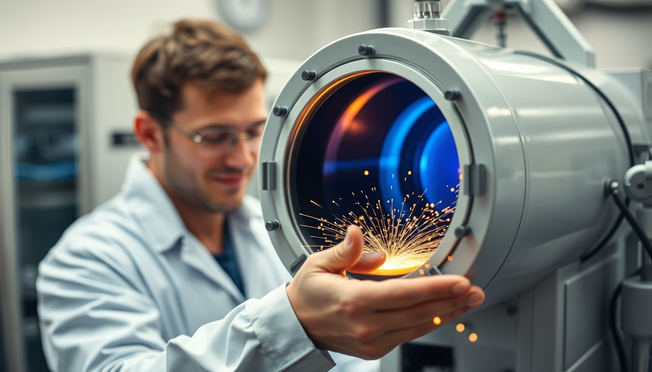
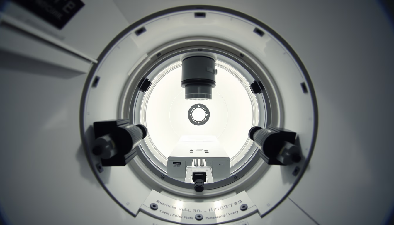
A câmara de microscópio eletrônico é o compartimento central onde a amostra é analisada sob feixe de elétrons em alto vácuo.
Sobre o tema
A câmara de um microscópio eletrônico é um componente crítico que abriga a amostra durante a análise. Diferente dos microscópios ópticos, que usam luz, os microscópios eletrônicos utilizam um feixe de elétrons para obter resoluções na escala nanométrica. Para que os elétrons possam percorrer seu trajeto sem serem desviados por moléculas de ar, a câmara é mantida em alto vácuo, normalmente na faixa de 10^-4 a 10^-7 Pa. Esse vácuo é obtido por uma combinação de bombas mecânicas e difusoras ou turbo-moleculares.
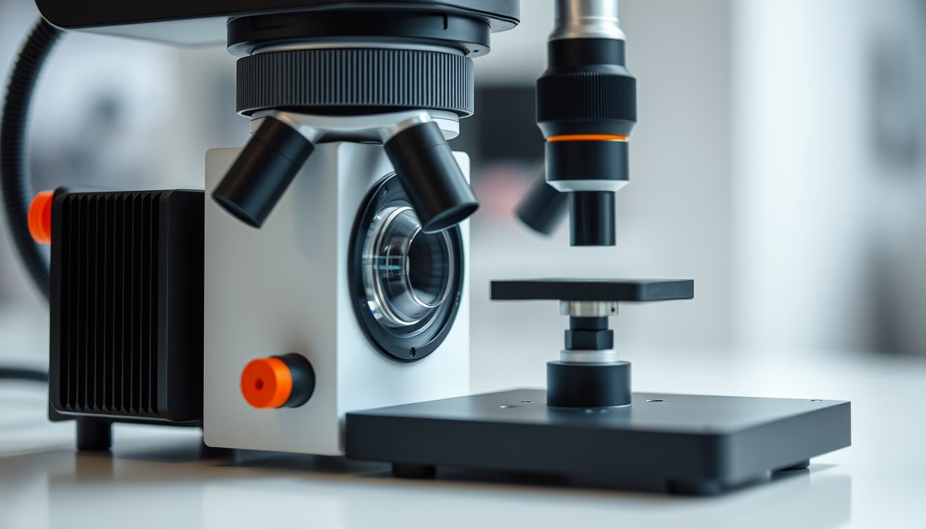
Existem dois tipos principais de microscópios eletrônicos: o de varredura (SEM) e o de transmissão (TEM). No SEM, a câmara é projetada para que o feixe varra a superfície da amostra, gerando elétrons secundários e retroespalhados que formam a imagem. No TEM, o feixe atravessa a amostra ultrafina, e a câmara deve ser mais profunda para acomodar o sistema de lentes e detectores abaixo. Ambas as câmaras possuem portas para inserção de amostras, janelas de visualização e múltiplos conectores para detectores e acessórios.
A preparação da amostra é essencial: materiais não condutores precisam ser revestidos com ouro ou carbono para evitar acúmulo de carga. Amostras biológicas passam por fixação, desidratação e metalização. A câmara também pode conter estágios motorizados que permitem movimentação precisa nos eixos X, Y, Z e inclinação, possibilitando a análise tridimensional. A manutenção do vácuo é tão rigorosa que qualquer vazamento mínimo pode comprometer a qualidade da imagem.
Curiosamente, as primeiras câmaras de microscópio eletrônico foram desenvolvidas na década de 1930 por Ernst Ruska e Max Knoll. Desde então, evoluíram para sistemas com controle computadorizado, automação de troca de amostras e integração com espectroscopia de raios X (EDS). A câmara não é apenas um receptáculo; ela é o ambiente onde a interação entre elétrons e matéria revela estruturas invisíveis a olho nu.
Perguntas frequentes
Por que a câmara do microscópio eletrônico precisa de vácuo?
O vácuo é necessário para que o feixe de elétrons não colida com moléculas de ar, o que desviaria os elétrons e reduziria a resolução. Além disso, evita a contaminação da amostra e dos componentes eletrônicos.
Qual a diferença entre a câmara de um SEM e de um TEM?
No SEM, a câmara é otimizada para que o feixe varra a superfície da amostra, geralmente com um volume maior para acomodar detectores de elétrons secundários. No TEM, a câmara é mais profunda e permite a passagem do feixe através da amostra, com espaço para lentes e detectores abaixo.
Como as amostras são preparadas para análise na câmara?
Amostras não condutoras recebem um revestimento condutivo (ouro, carbono) para evitar cargas elétricas. Amostras biológicas são fixadas quimicamente, desidratadas e metalizadas. Amostras para TEM devem ser ultrafinas (cerca de 100 nm) para permitir a transmissão de elétrons.
URL direta
https://pub-c7d6a6ea828543ac903a74a341ccb2e1.r2.dev/imagens/electron-microscope-chamber-fine-art-photography-fresh6.avifComo creditar
Inclua um link visível de volta pro UtilizAí. Copie um dos snippets abaixo:
<a href="https://xn--utiliza-eza.com/midia/imagens/electron-microscope-chamber-fine-art-photography-fresh6">Electron microscope chamber</a> by <a href="https://xn--utiliza-eza.com">UtilizAí</a>, licensed under <a href="https://creativecommons.org/licenses/by/4.0/">CC BY 4.0</a>.
[Electron microscope chamber](https://xn--utiliza-eza.com/midia/imagens/electron-microscope-chamber-fine-art-photography-fresh6) by [UtilizAí](https://xn--utiliza-eza.com), CC BY 4.0
Licença: CC-BY-4.0