Electron microscope chamber
1344×768 · AVIF · CC BY 4.0
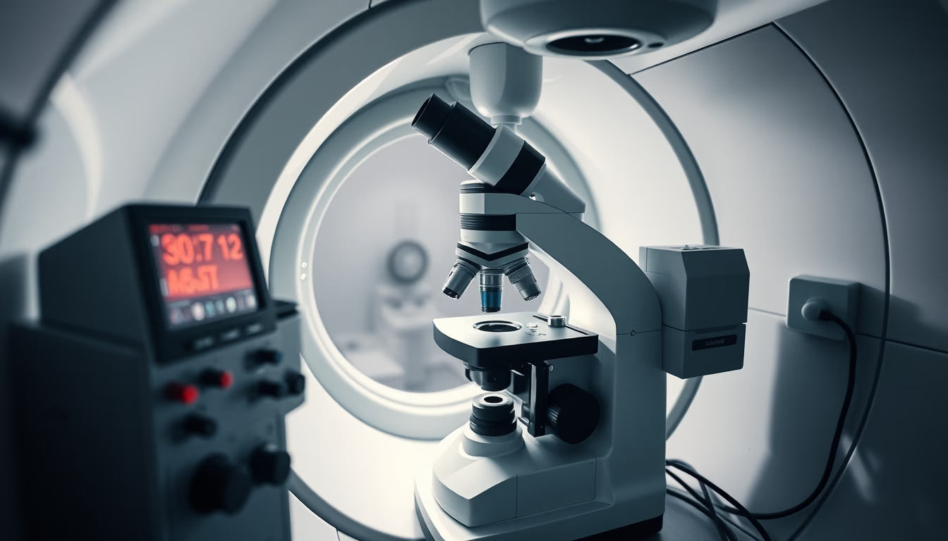
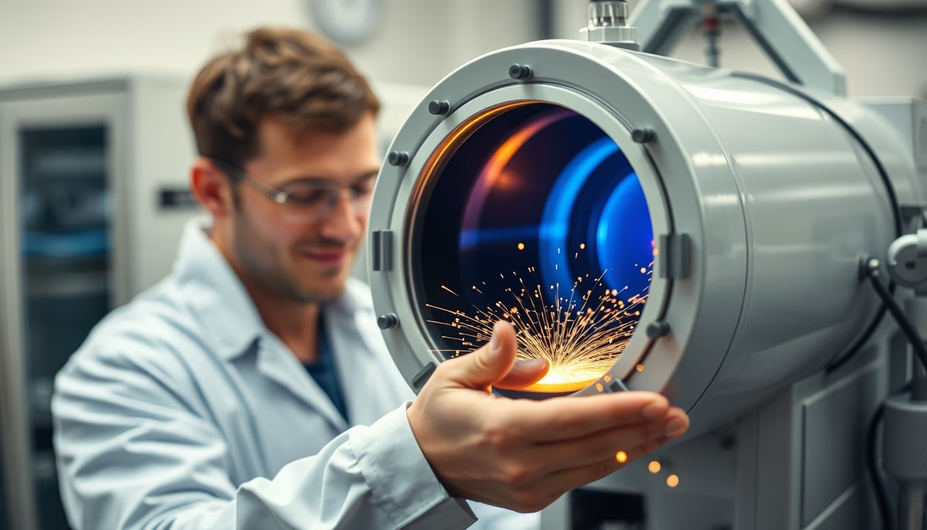
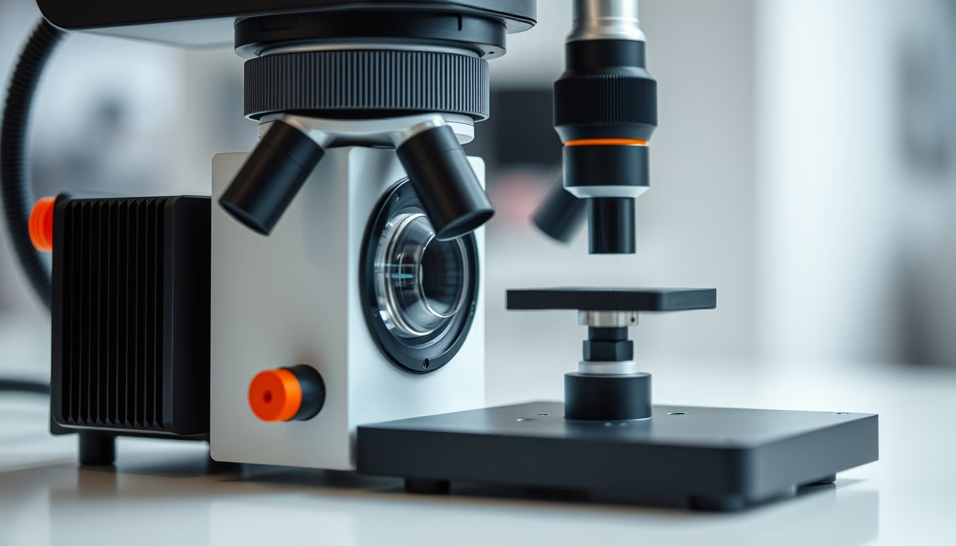
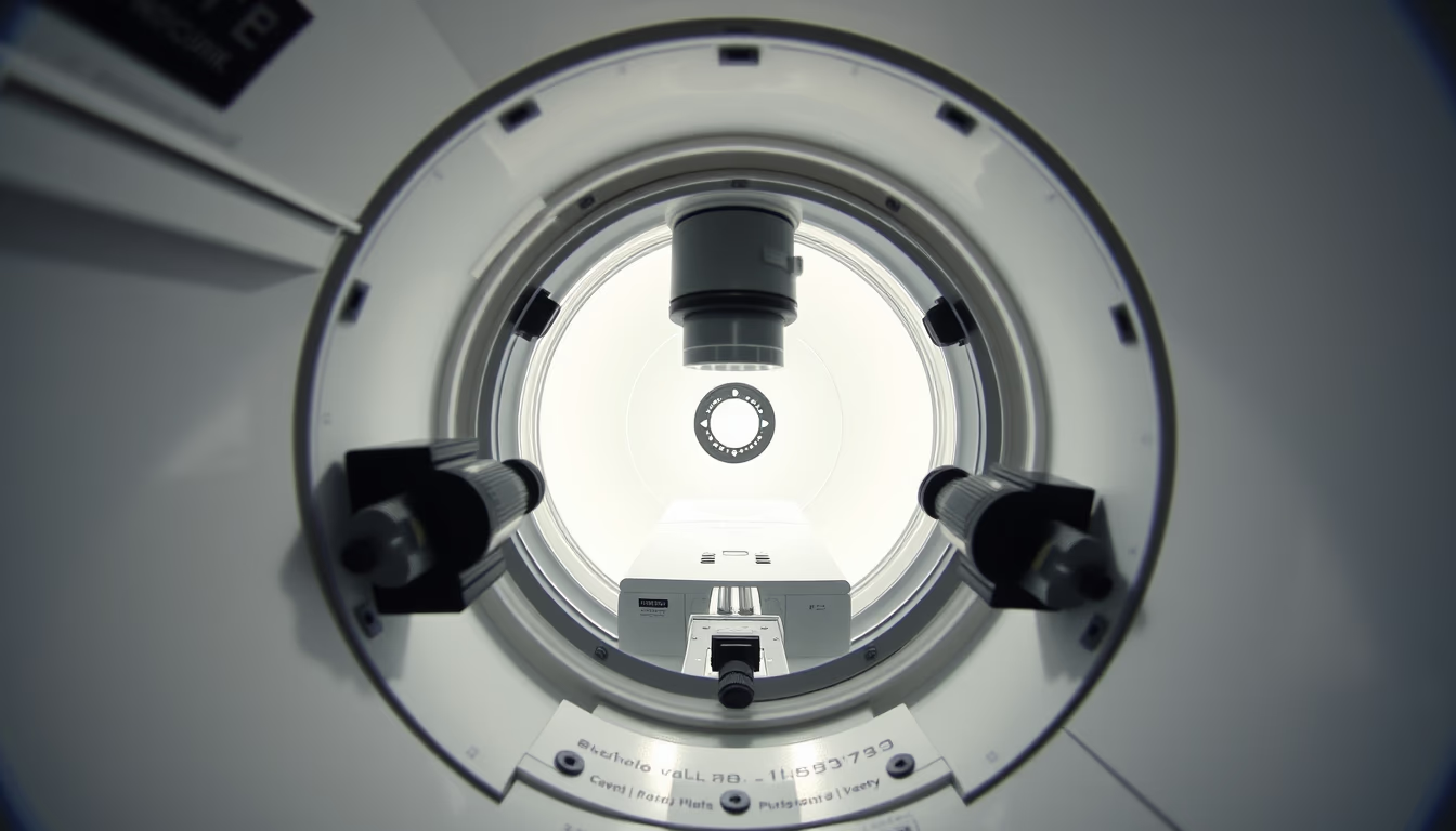
The chamber of an electron microscope is the heart of the equipment, where electron beams interact with the sample under high vacuum to generate nanoscale images.
About this subject
The vacuum chamber of an electron microscope is one of the most critical components of the instrument. Inside, the sample is positioned and exposed to a focused electron beam. The vacuum is essential to prevent air molecules from scattering the electrons, allowing resolution down to individual atoms. Scanning electron microscopes (SEM) and transmission electron microscopes (TEM) operate at pressures around 10⁻⁴ to 10⁻⁷ Pa.
Historically, the first practical electron microscope was built by Ernst Ruska and Max Knoll in 1931 in Germany. Since then, chambers have evolved significantly: materials like stainless steel and special alloys minimize magnetic interference, and turbomolecular and ion pumps maintain the necessary high vacuum. The chamber also houses detectors, manipulable sample stages, and in some models, cryogenic systems for observing sensitive materials.
Notably, chamber design directly impacts image quality. External electromagnetic interference is blocked by mu-metal shielding on the walls. Modern chambers allow the introduction of reactive gases for in situ experiments, such as oxidation or catalysis, expanding applications in materials science and biology.
In Brazil, electron microscopes in universities and research centers are essential for nanotechnology, metallurgy, and virology. Operating and maintaining these chambers requires specialized technicians, as any contamination or vacuum loss can compromise days of experimentation.
Frequently Asked Questions
Why does the electron microscope chamber need a vacuum?
The vacuum prevents air molecules from deflecting or scattering the electron beam, allowing it to hit the sample precisely and produce high-resolution images. Without vacuum, electrons would collide with air particles, drastically reducing sharpness.
What is the difference between an SEM and a TEM chamber?
In a scanning electron microscope (SEM), the chamber is larger to accommodate bulky samples and secondary electron detectors. In a transmission electron microscope (TEM), the chamber is more compact and includes a lens system to focus the beam on the ultrathin sample.
How is the vacuum chamber maintained?
Maintenance involves periodic cleaning with special solvents, replacing sealing gaskets, and checking vacuum pumps. Trained technicians perform leak tests and calibration to ensure stable pressures.
Direct URL
https://pub-c7d6a6ea828543ac903a74a341ccb2e1.r2.dev/imagens/electron-microscope-chamber-magazine-editorial-fresh7.avifHow to credit
Include a visible link back to UtilizAí. Copy one of the snippets below:
<a href="https://xn--utiliza-eza.com/en/midia/imagens/electron-microscope-chamber-magazine-editorial-fresh7">Electron microscope chamber</a> by <a href="https://xn--utiliza-eza.com">UtilizAí</a>, licensed under <a href="https://creativecommons.org/licenses/by/4.0/">CC BY 4.0</a>.
[Electron microscope chamber](https://xn--utiliza-eza.com/en/midia/imagens/electron-microscope-chamber-magazine-editorial-fresh7) by [UtilizAí](https://xn--utiliza-eza.com), CC BY 4.0
License: CC-BY-4.0