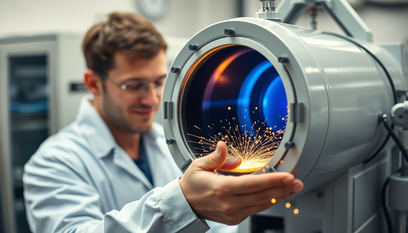
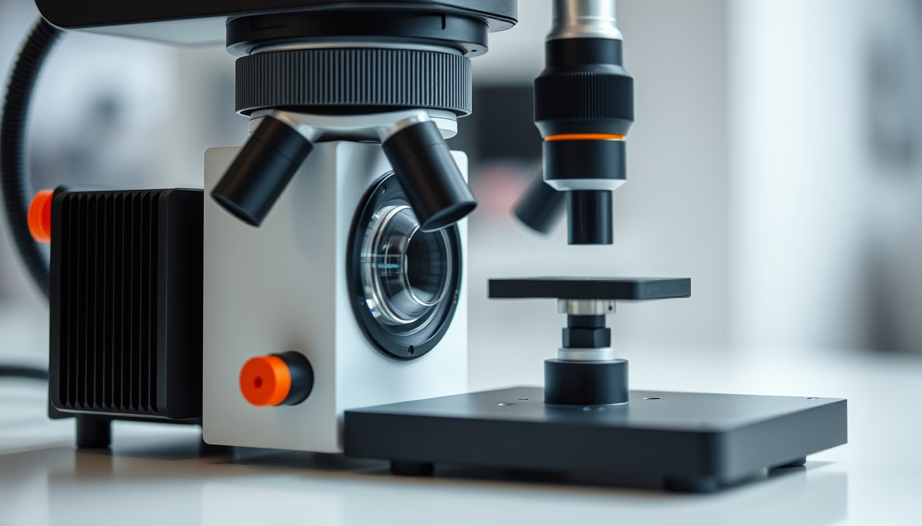
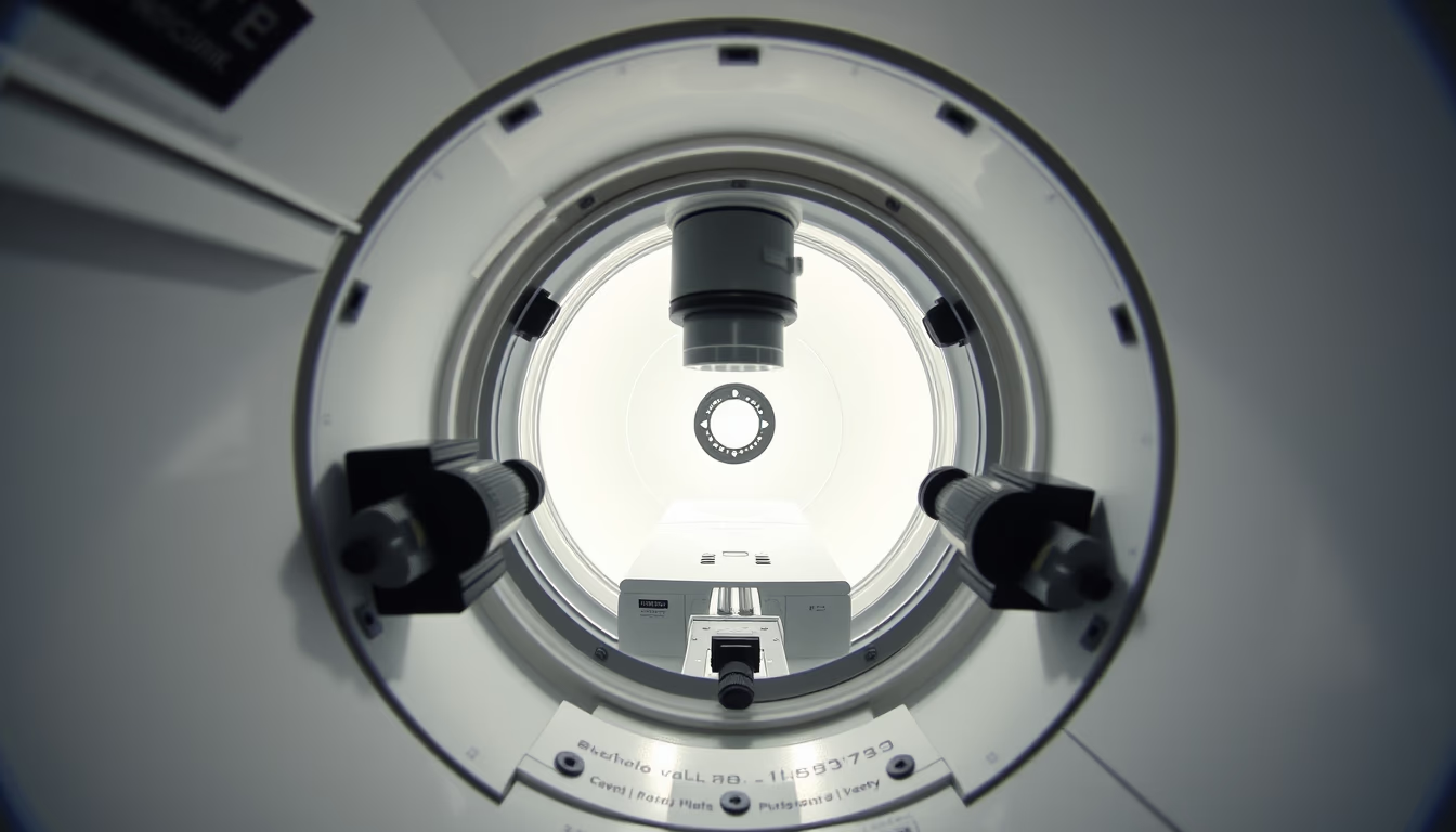
Electron microscope chamber
1344×768 · AVIF · CC BY 4.0
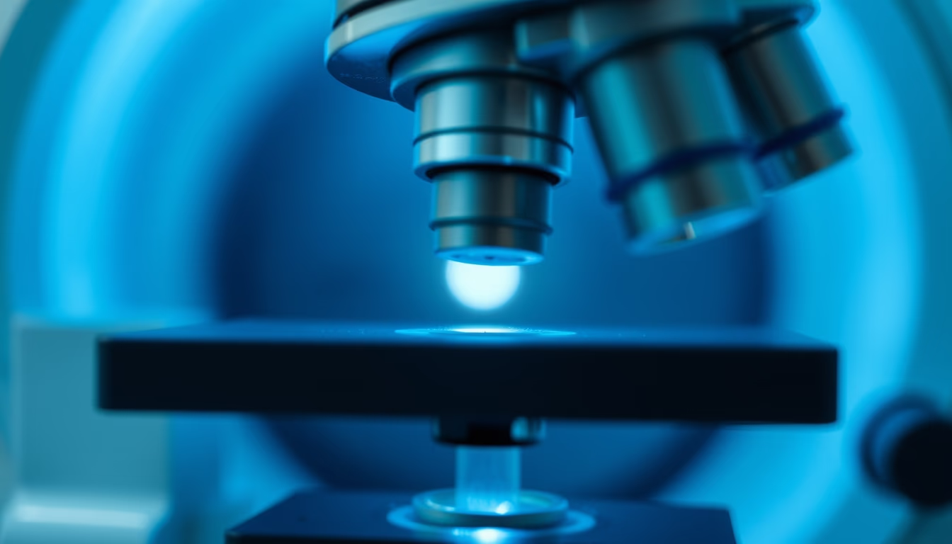
The electron microscope chamber is the core of the instrument, where electron beams interact with samples in a vacuum, producing images at nanometer scale.
About this subject
The electron microscope chamber is a sealed environment where the sample is placed for analysis. It maintains an ultra-high vacuum (10⁻⁴ to 10⁻⁷ Pa) to prevent air molecules from scattering the electron beam. Inside, electromagnetic lenses, detectors, and precision stages allow movement in x, y, z, and rotation axes. The vacuum also preserves sample integrity, especially for sensitive materials.
The electron beam, generated by a tungsten filament or field emission gun, is accelerated at 1, 300 kV and focused by the lenses. Upon hitting the sample, electrons produce signals such as secondary and backscattered electrons, captured by specific detectors. These signals are converted into images with magnifications up to 10 million times, revealing details as small as a few nanometers.
The chamber is crucial in fields like cell biology (visualizing viruses and organelles), materials science (failure analysis of metals), and nanotechnology (characterization of nanoparticles). Unlike optical microscopes, electron microscopes overcome the diffraction limit, achieving atomic resolution. Modern instruments incorporate chambers with multiple detectors for elemental analysis (EDS) and diffraction (EBSD).
Maintaining the chamber requires rigorous care: avoiding vibrations, external magnetic fields, and contamination. Pumping systems (mechanical, turbomolecular, and ion pumps) ensure the vacuum. The high cost and need for controlled environments restrict these instruments to specialized laboratories.
Frequently Asked Questions
What is the purpose of the vacuum chamber in an electron microscope?
The vacuum chamber removes air molecules that could scatter the electron beam, enabling clear, high-resolution images. Without a vacuum, electrons would collide with air atoms, losing energy and degrading image quality.
What is the difference between SEM and TEM regarding the chamber?
In a scanning electron microscope (SEM), the sample is in a separate chamber and the beam scans the surface, producing 3D-like images. In a transmission electron microscope (TEM), the sample is ultra-thin and the beam passes through, with detectors below, requiring an even higher vacuum.
What materials are used to build an electron microscope chamber?
The chamber is typically made of stainless steel or special alloys to resist corrosion and maintain vacuum. Beryllium or polymer windows allow radiation passage, and elastomer or metal seals ensure airtightness.
Direct URL
https://pub-c7d6a6ea828543ac903a74a341ccb2e1.r2.dev/imagens/electron-microscope-chamber-macro-closeup-fresh4.avifHow to credit
Include a visible link back to UtilizAí. Copy one of the snippets below:
<a href="https://xn--utiliza-eza.com/en/midia/imagens/electron-microscope-chamber-macro-closeup-fresh4">Electron microscope chamber</a> by <a href="https://xn--utiliza-eza.com">UtilizAí</a>, licensed under <a href="https://creativecommons.org/licenses/by/4.0/">CC BY 4.0</a>.
[Electron microscope chamber](https://xn--utiliza-eza.com/en/midia/imagens/electron-microscope-chamber-macro-closeup-fresh4) by [UtilizAí](https://xn--utiliza-eza.com), CC BY 4.0
License: CC-BY-4.0