Electron microscope chamber
1344×768 · AVIF · CC BY 4.0
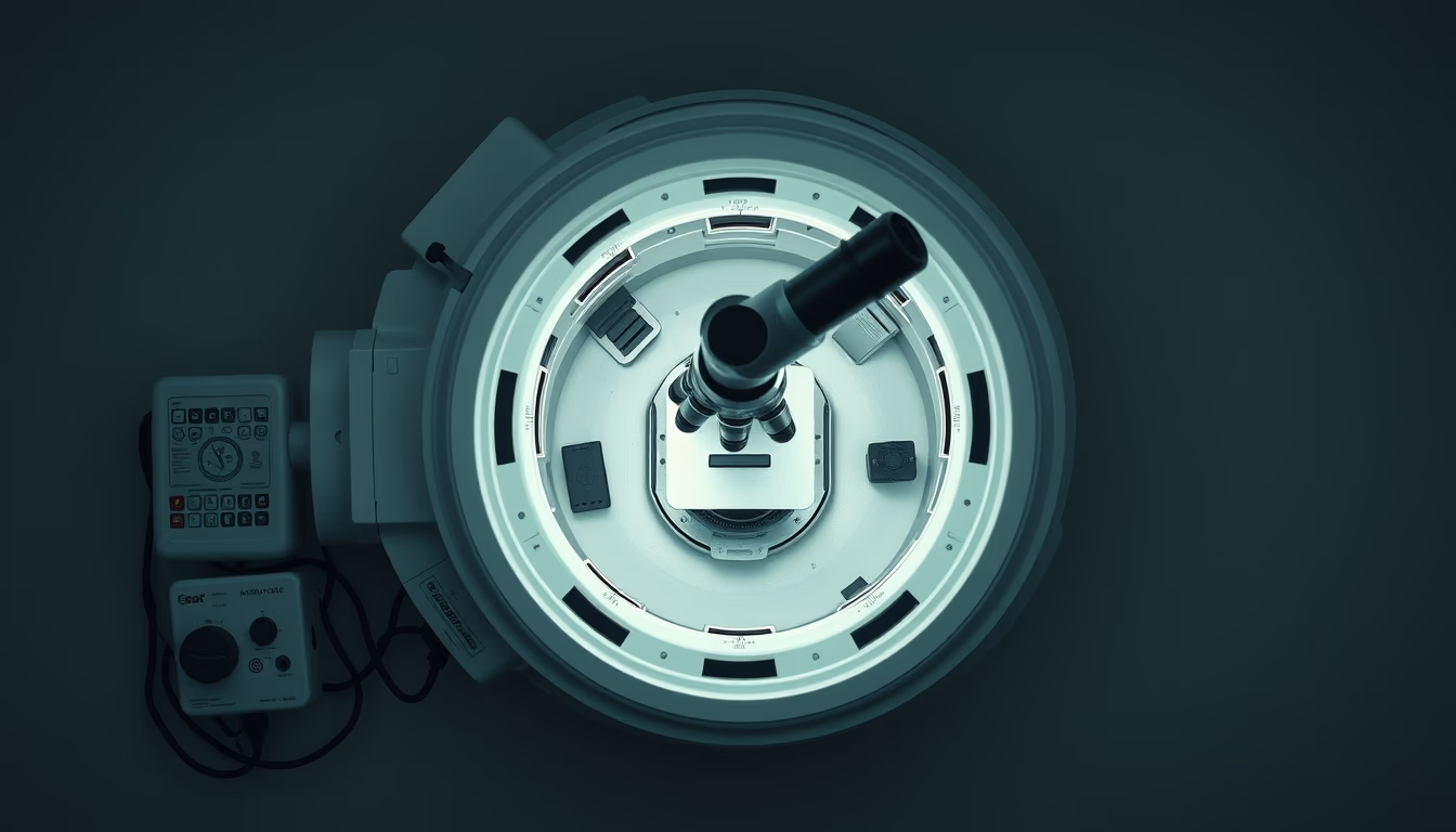
The chamber of an electron microscope is the core component where electron beams interact with the sample under vacuum to produce nanoscale images.
About this subject
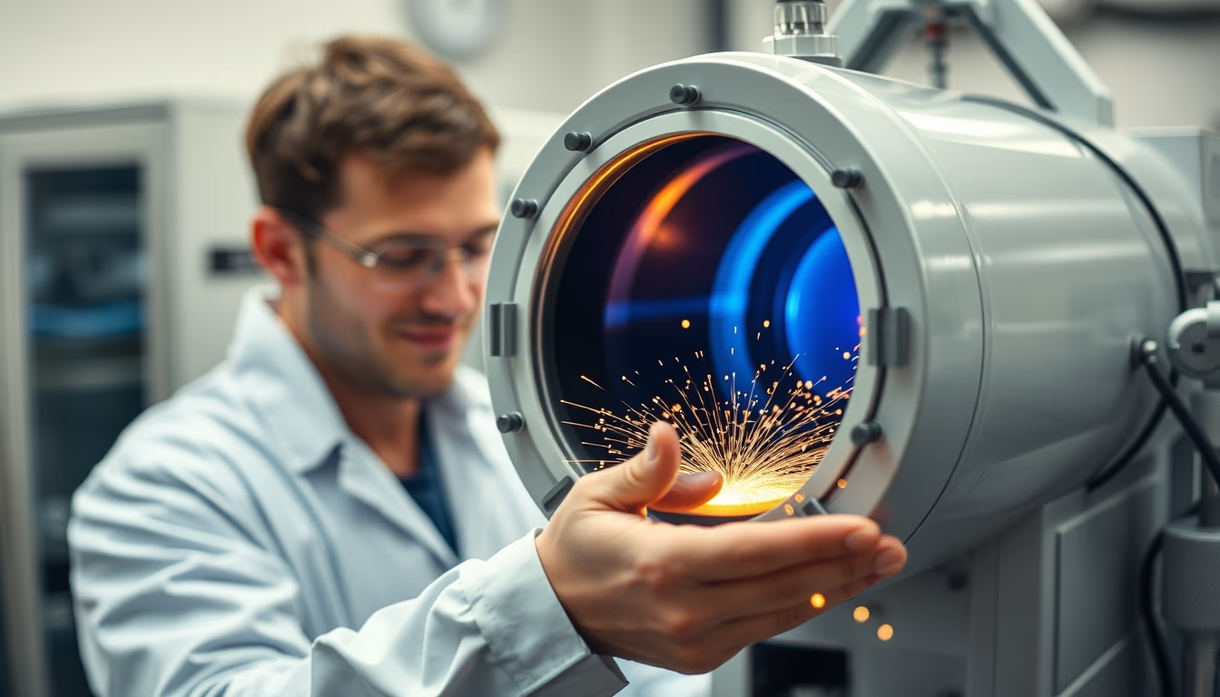
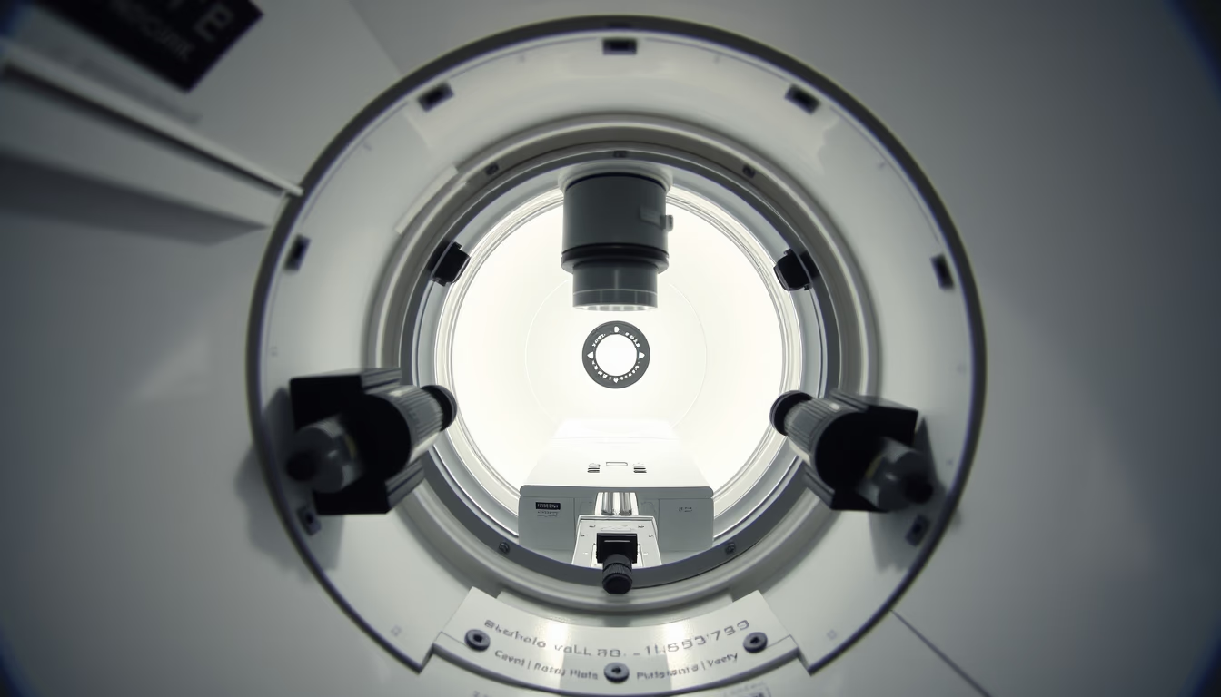
The vacuum chamber of an electron microscope is a sealed compartment that houses the sample and detection systems during analysis. Unlike optical microscopes that use light, electron microscopes employ beams of electrons accelerated by electromagnetic fields. For these electrons to travel without colliding with air molecules, the chamber is maintained at high vacuum, typically in the range of 10^-4 to 10^-7 Pascal. This controlled environment is essential to prevent electron scattering and ensure imaging resolution down to 0.1 nanometers, enough to visualize individual atoms.
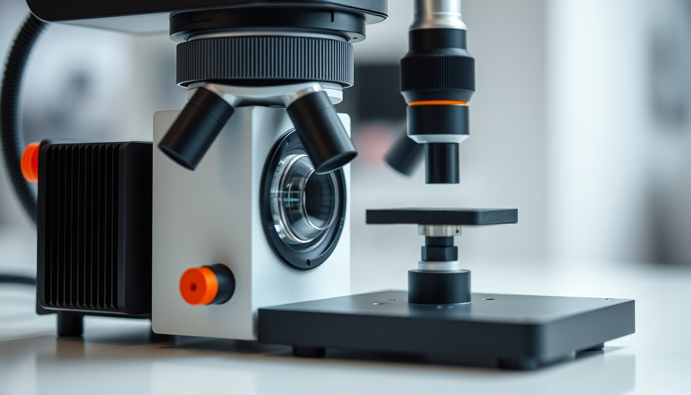
There are two main types of electron microscopes: scanning (SEM) and transmission (TEM). In SEM, the beam scans the sample surface, and secondary electrons produce topographical images. In TEM, electrons pass through an ultrathin sample, generating images of internal structure. In both, the chamber houses detectors, electromagnetic lenses, and specimen holders. The vacuum system includes mechanical and diffusion pumps, which remove gases and vapors, as well as cryogenic traps to capture impurities.
Historically, the first electron microscope was built by Ernst Ruska in the 1930s, work that earned him the Nobel Prize in Physics in 1986. Since then, technology has evolved with more efficient vacuum systems, thermal stability, and spherical aberration correction. Modern electron microscope chambers include features such as low residual gas, quick-access ports, and interfaces for in situ manipulation, allowing observation of chemical reactions or temperature changes in real time.
The applications of these instruments are vast: from materials science and nanotechnology to structural biology and semiconductors. In microelectronics, for instance, the vacuum chamber is used to inspect defects in computer chips. In medicine, cryo-electron microscopy (cryo-EM) revolutionized the study of proteins and viruses, earning the Nobel Prize in Chemistry in 2017. Thus, the vacuum chamber, although just a part of the system, is a critical element that enables observation of the world invisible to the naked eye.
Frequently Asked Questions
What is the function of the vacuum chamber in an electron microscope?
The vacuum chamber maintains an ultra-low pressure environment to prevent electrons from colliding with air molecules, ensuring a clear path and atomic-scale imaging.
What types of pumps are used to create vacuum in these microscopes?
Mechanical pumps (for rough vacuum) and diffusion or turbomolecular pumps (for high vacuum) are used, along with cryogenic traps to remove traces of water and hydrocarbons.
Why is vacuum more critical in transmission electron microscopy (TEM) than in scanning (SEM)?
In TEM, electrons must pass through the sample and travel a long column; any gas collisions degrade the image. In SEM, while still important, vacuum can be slightly less stringent (10^-3 Pa) because electrons do not need to traverse the sample.
Direct URL
https://pub-c7d6a6ea828543ac903a74a341ccb2e1.r2.dev/imagens/electron-microscope-chamber-isometric-flat-lay-fresh16.avifHow to credit
Include a visible link back to UtilizAí. Copy one of the snippets below:
<a href="https://xn--utiliza-eza.com/en/midia/imagens/electron-microscope-chamber-isometric-flat-lay-fresh16">Electron microscope chamber</a> by <a href="https://xn--utiliza-eza.com">UtilizAí</a>, licensed under <a href="https://creativecommons.org/licenses/by/4.0/">CC BY 4.0</a>.
[Electron microscope chamber](https://xn--utiliza-eza.com/en/midia/imagens/electron-microscope-chamber-isometric-flat-lay-fresh16) by [UtilizAí](https://xn--utiliza-eza.com), CC BY 4.0
License: CC-BY-4.0