Electron microscope chamber
1344×768 · AVIF · CC BY 4.0

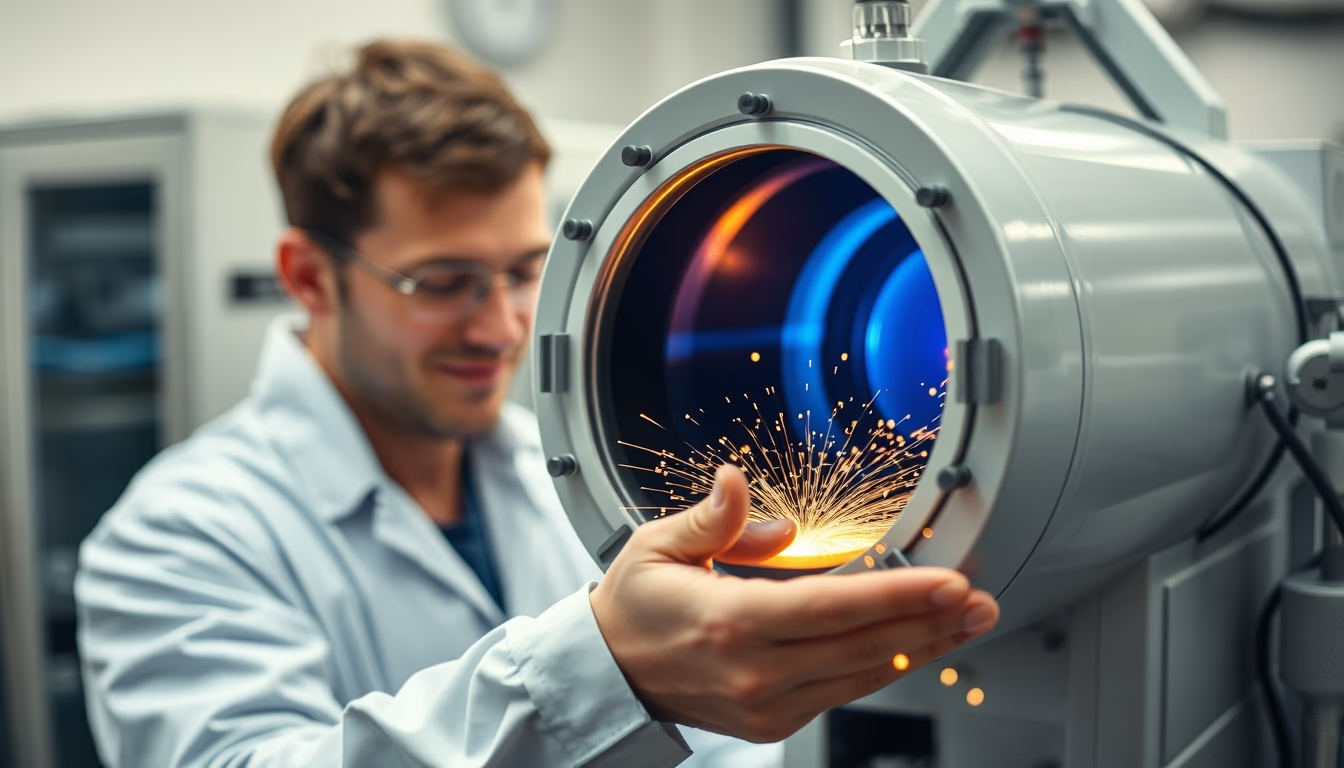
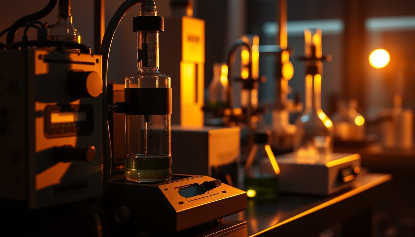
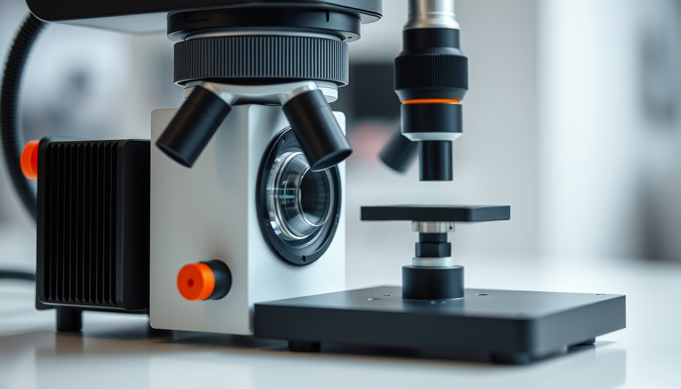
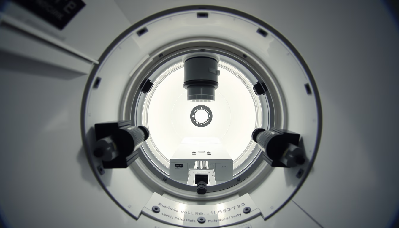
The electron microscope chamber is the vacuum-sealed compartment where the sample is analyzed by an electron beam, essential for achieving magnifications up to 2 million times.
About this subject
The electron microscope chamber is a central component in both scanning electron microscopes (SEM) and transmission electron microscopes (TEM). Its main function is to maintain a high-vacuum environment (typically between 10^-4 and 10^-8 Pa) to prevent air molecules from scattering the electron beam, ensuring a clear path and sharp images. Inside the chamber, the sample is mounted on a stage with precise movement along X, Y, Z axes and rotation, enabling analysis from various angles.
The development of vacuum chambers dates back to the 1930s when Ernst Ruska built the first electron microscope, for which he received the Nobel Prize in Physics in 1986. Since then, materials and engineering have advanced; modern chambers are made of stainless steel or aluminum with beryllium windows for detectors. In TEM, the chamber also houses electromagnetic lenses that focus the beam and a detection system that captures transmitted electrons, forming images with sub-nanometer resolution.
The chamber's versatility allows the integration of various detectors, such as EDX for elemental analysis and EBSD for crystallography. Additionally, accessories like heating or cooling stages (down to -190 °C with liquid nitrogen) and mechanical deformation holders expand applications. Environmental SEM (ESEM) technology enables imaging of wet or biological samples without full dehydration, revolutionizing fields like cell biology.
Chamber maintenance is critical: vacuum is achieved by a system of mechanical and diffusion or turbomolecular pumps, and any leak can compromise both the image and the beam. Sample preparation techniques, such as sputter coating with gold or carbon, are performed outside the chamber, but evacuation time can take minutes. The electron microscope chamber is thus a microcosm of precision engineering that enables exploration of the invisible world.
Frequently Asked Questions
Why does the electron microscope chamber need a vacuum?
The vacuum prevents air molecules from scattering or deflecting the electron beam, which would degrade image resolution. It also protects the filament (electron source) from oxidation and allows electrons to travel in a straight line to the detector.
What is the difference between the chamber of an SEM and a TEM?
In an SEM, the chamber is larger and the sample is tilted, detecting secondary electrons reflected from the surface. In a TEM, the chamber is elongated, the sample is ultrathin (less than 100 nm), and electrons pass through it, forming an image by density contrast. Both use vacuum, but TEM requires even higher vacuum to avoid contamination.
What samples can be analyzed in an electron microscope chamber?
Conductive or metal-coated samples (for SEM) and ultrathin sections (for TEM). Biological materials must be fixed, dehydrated, and coated. Environmental SEM (ESEM) allows wet samples but with lower resolution. Rocks, metals, ceramics, polymers, and nanostructures are common examples.
Direct URL
https://pub-c7d6a6ea828543ac903a74a341ccb2e1.r2.dev/imagens/electron-microscope-chamber-high-fashion-editorial-fresh10.avifHow to credit
Include a visible link back to UtilizAí. Copy one of the snippets below:
<a href="https://xn--utiliza-eza.com/en/midia/imagens/electron-microscope-chamber-high-fashion-editorial-fresh10">Electron microscope chamber</a> by <a href="https://xn--utiliza-eza.com">UtilizAí</a>, licensed under <a href="https://creativecommons.org/licenses/by/4.0/">CC BY 4.0</a>.
[Electron microscope chamber](https://xn--utiliza-eza.com/en/midia/imagens/electron-microscope-chamber-high-fashion-editorial-fresh10) by [UtilizAí](https://xn--utiliza-eza.com), CC BY 4.0
License: CC-BY-4.0