Electron microscope chamber
1344×768 · AVIF · CC BY 4.0
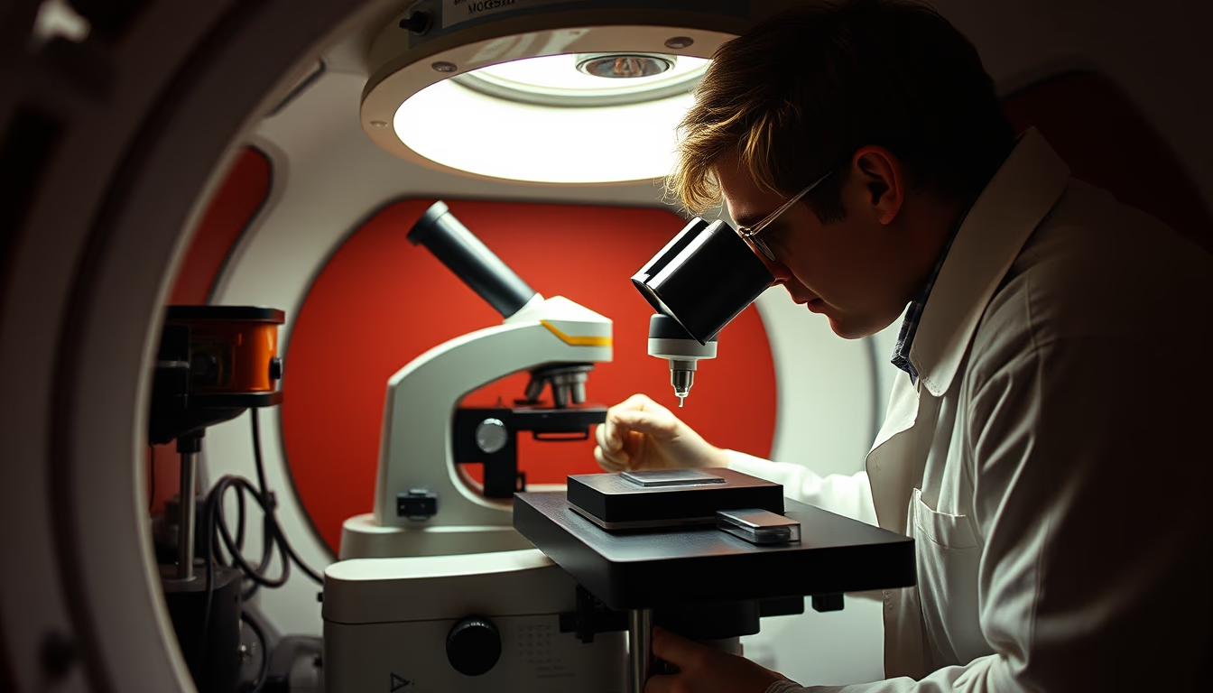
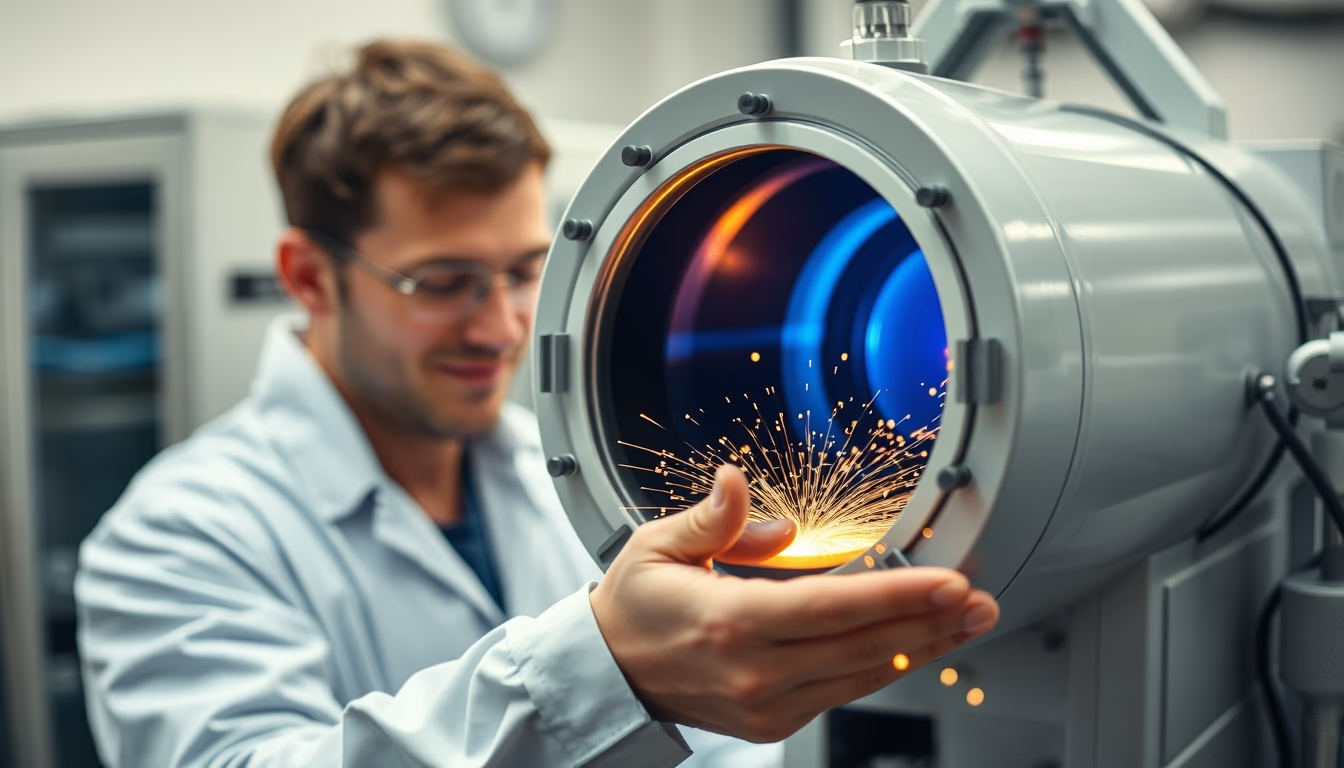
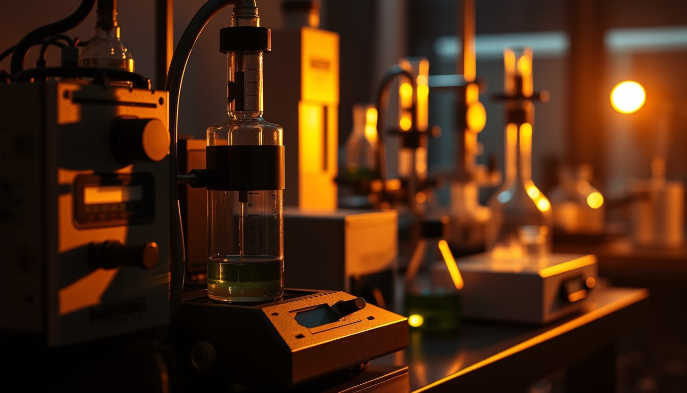
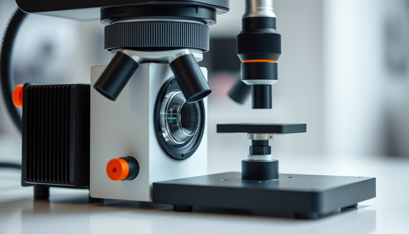
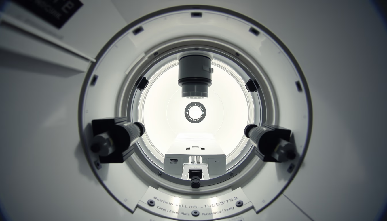
The electron microscope chamber is the core component where the electron beam interacts with the sample under vacuum to produce ultra-high-resolution images.
About this subject
The vacuum chamber of an electron microscope houses the sample and the lens system that focuses the electron beam. This sealed environment is essential to prevent air molecules from scattering the electrons, ensuring precise trajectories. Inside the chamber, the sample is placed on a movable stage that allows tilting and rotation. Specialized detectors convert signals from the electron-sample interaction into digital images.
There are two main types: transmission electron microscopes (TEM) and scanning electron microscopes (SEM). In TEM, electrons pass through an ultrathin sample to form a two-dimensional projection. In SEM, the beam scans the sample surface, producing three-dimensional images with high depth of field. Both require high vacuum levels (10^-4 to 10^-8 Pa) to avoid contamination and resolution loss.
Invented by Ernst Ruska and Max Knoll in 1931, the electron microscope enabled visualization at the atomic scale. Modern chambers incorporate cryogenic systems for biological samples, rapid load locks, and high-precision motorized stages. Applications include semiconductor failure analysis, nanomaterial characterization, and studying viruses like SARS-CoV-2.
Maintaining the chamber free of vibrations and external magnetic fields is critical. Many microscopes are mounted on vibration isolation tables and installed in shielded rooms. Periodic chamber cleaning and the use of transfer chambers minimize impurity introduction.
Frequently Asked Questions
Why does the electron microscope chamber need vacuum?
Vacuum prevents air molecules from scattering or deflecting electrons, which would degrade image resolution. It also avoids contamination of the sample and lens system by particles or vapors.
What is the difference between TEM and SEM chambers?
In TEM, the sample is thin and electrons pass through, requiring precise beam alignment. In SEM, the surface is scanned, needing secondary electron detectors. Both use vacuum, but TEM typically requires higher (ultra-high) vacuum.
How are samples prepared for electron microscope observation?
For TEM, samples must be ultrathin (under 100 nm), cut with a microtome or focused ion beam. For SEM, larger blocks are possible but require conductive coating (gold, carbon) if non-conductive. Both must be dry and stable under vacuum.
Direct URL
https://pub-c7d6a6ea828543ac903a74a341ccb2e1.r2.dev/imagens/electron-microscope-chamber-film-grain-authentic-fresh12.avifHow to credit
Include a visible link back to UtilizAí. Copy one of the snippets below:
<a href="https://xn--utiliza-eza.com/en/midia/imagens/electron-microscope-chamber-film-grain-authentic-fresh12">Electron microscope chamber</a> by <a href="https://xn--utiliza-eza.com">UtilizAí</a>, licensed under <a href="https://creativecommons.org/licenses/by/4.0/">CC BY 4.0</a>.
[Electron microscope chamber](https://xn--utiliza-eza.com/en/midia/imagens/electron-microscope-chamber-film-grain-authentic-fresh12) by [UtilizAí](https://xn--utiliza-eza.com), CC BY 4.0
License: CC-BY-4.0