Electron microscope chamber
1344×768 · AVIF · CC BY 4.0
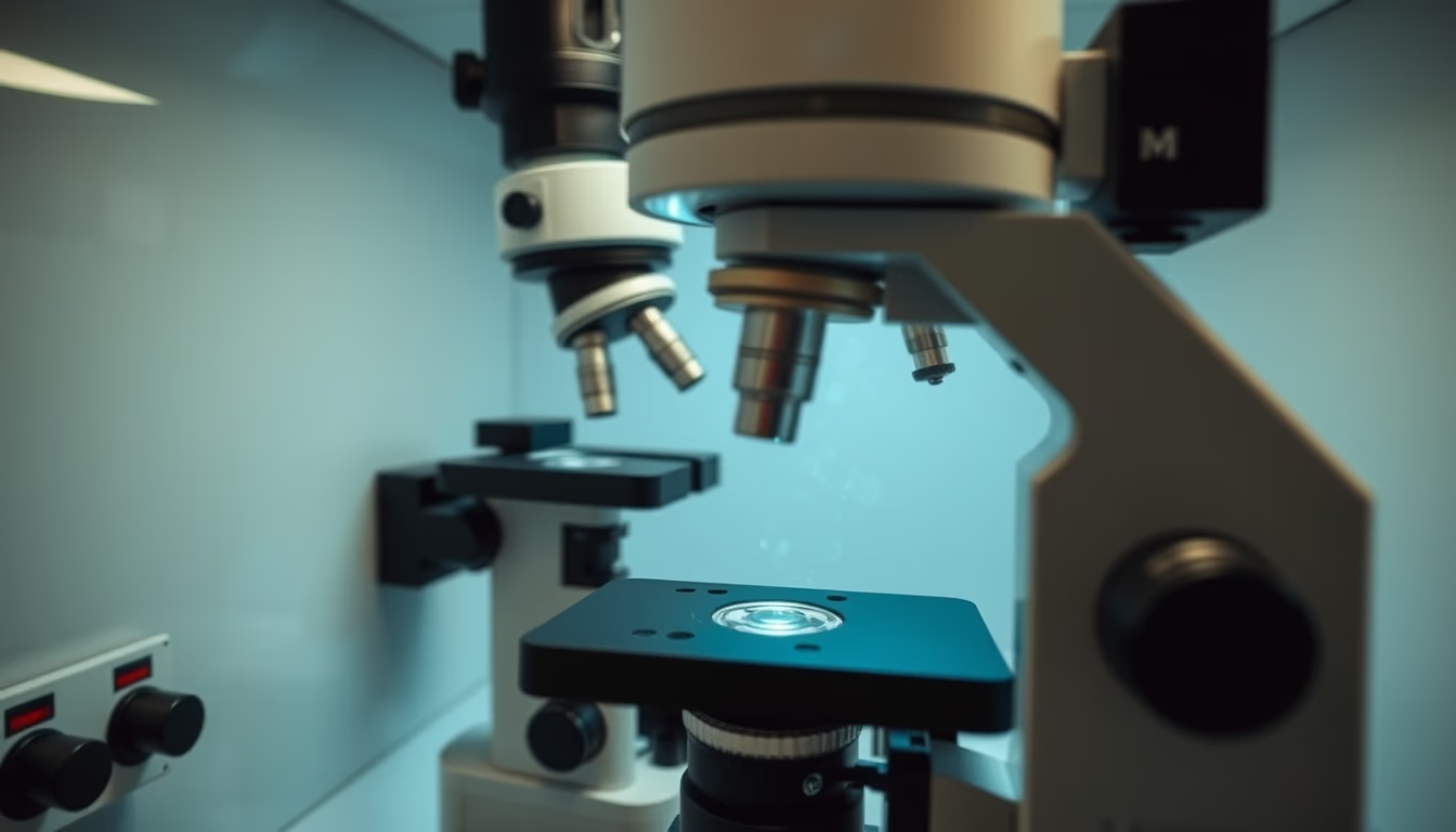
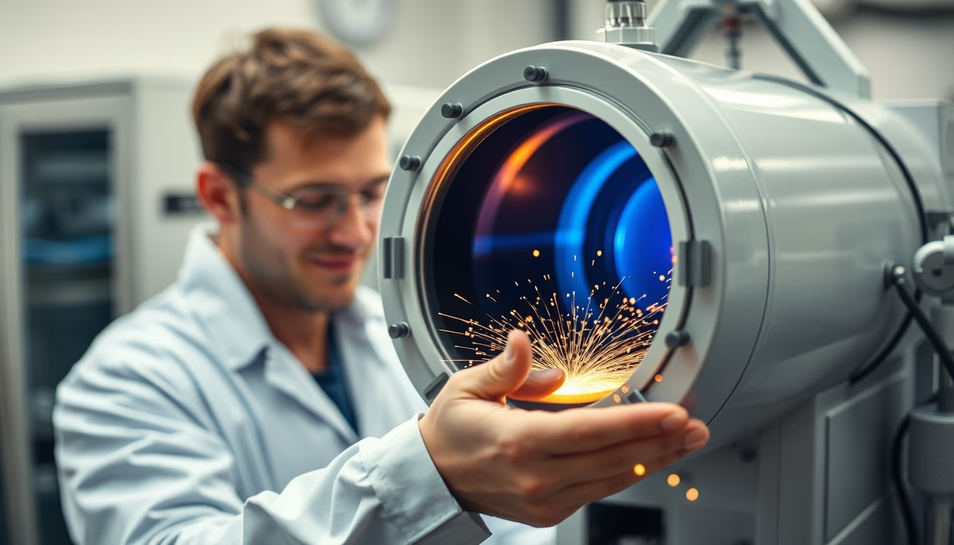
The chamber of an electron microscope is the core component where electron beams interact with the sample under high vacuum conditions.
About this subject
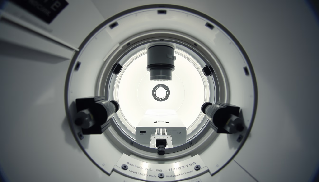
The vacuum chamber of an electron microscope is a sealed compartment designed to maintain high vacuum conditions, essential for the electron beam to function. Inside, electrons are accelerated and focused by electromagnetic lenses, allowing imaging with nanometer resolution. Unlike optical microscopes that use visible light, electron microscopes use electrons to overcome the diffraction limit of light, enabling magnifications of up to 10 million times.
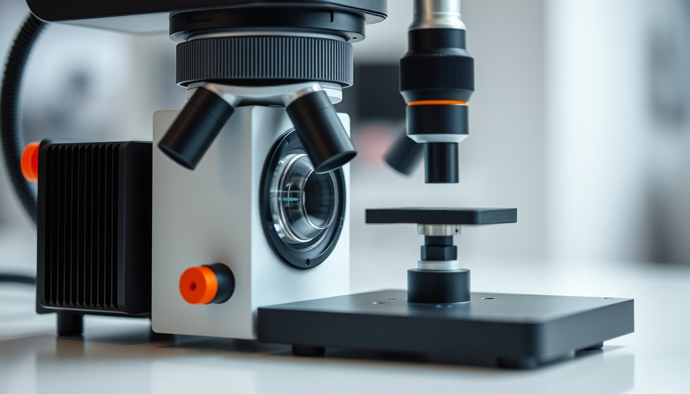
Within the chamber, the sample is placed on a specimen holder that can be tilted, heated, or cooled depending on the technique. Vacuum is necessary because air molecules would scatter the electrons, degrading the image. Vacuum pumps, such as turbomolecular and ion pumps, maintain internal pressure at extremely low levels, around 10⁻⁴ Pa or less. There are two main types of electron microscopes: the scanning electron microscope (SEM), which examines sample surfaces, and the transmission electron microscope (TEM), which analyzes the internal structure of ultrathin materials.
The invention of the electron microscope revolutionized science. The first prototype was built in 1931 by Ernst Ruska and Max Knoll in Germany, work that earned Ruska the Nobel Prize in Physics in 1986. Today, these instruments are indispensable in fields such as nanotechnology, cell biology, materials science, and semiconductors. The chamber may contain X-ray detectors for elemental analysis, CCD cameras for image capture, and cryogenic systems to preserve biological samples in their native state.
Frequently Asked Questions
What is the function of the vacuum chamber in an electron microscope?
The vacuum chamber creates an air-free environment so that electrons can travel without colliding with gas molecules, enabling focused beams and high-resolution imaging.
What are the main types of electron microscopes and how do they differ?
The main types are the scanning electron microscope (SEM), which images the sample surface, and the transmission electron microscope (TEM), which analyzes internal structure using electrons that pass through the sample.
When were electron microscopes invented and by whom?
The first electron microscope was built in 1931 by German physicists Ernst Ruska and Max Knoll. Ruska was awarded the Nobel Prize in 1986 for this contribution.
Direct URL
https://pub-c7d6a6ea828543ac903a74a341ccb2e1.r2.dev/imagens/electron-microscope-chamber-documentary-nature-style-fresh9.avifHow to credit
Include a visible link back to UtilizAí. Copy one of the snippets below:
<a href="https://xn--utiliza-eza.com/en/midia/imagens/electron-microscope-chamber-documentary-nature-style-fresh9">Electron microscope chamber</a> by <a href="https://xn--utiliza-eza.com">UtilizAí</a>, licensed under <a href="https://creativecommons.org/licenses/by/4.0/">CC BY 4.0</a>.
[Electron microscope chamber](https://xn--utiliza-eza.com/en/midia/imagens/electron-microscope-chamber-documentary-nature-style-fresh9) by [UtilizAí](https://xn--utiliza-eza.com), CC BY 4.0
License: CC-BY-4.0