Electron microscope chamber
1344×768 · AVIF · CC BY 4.0
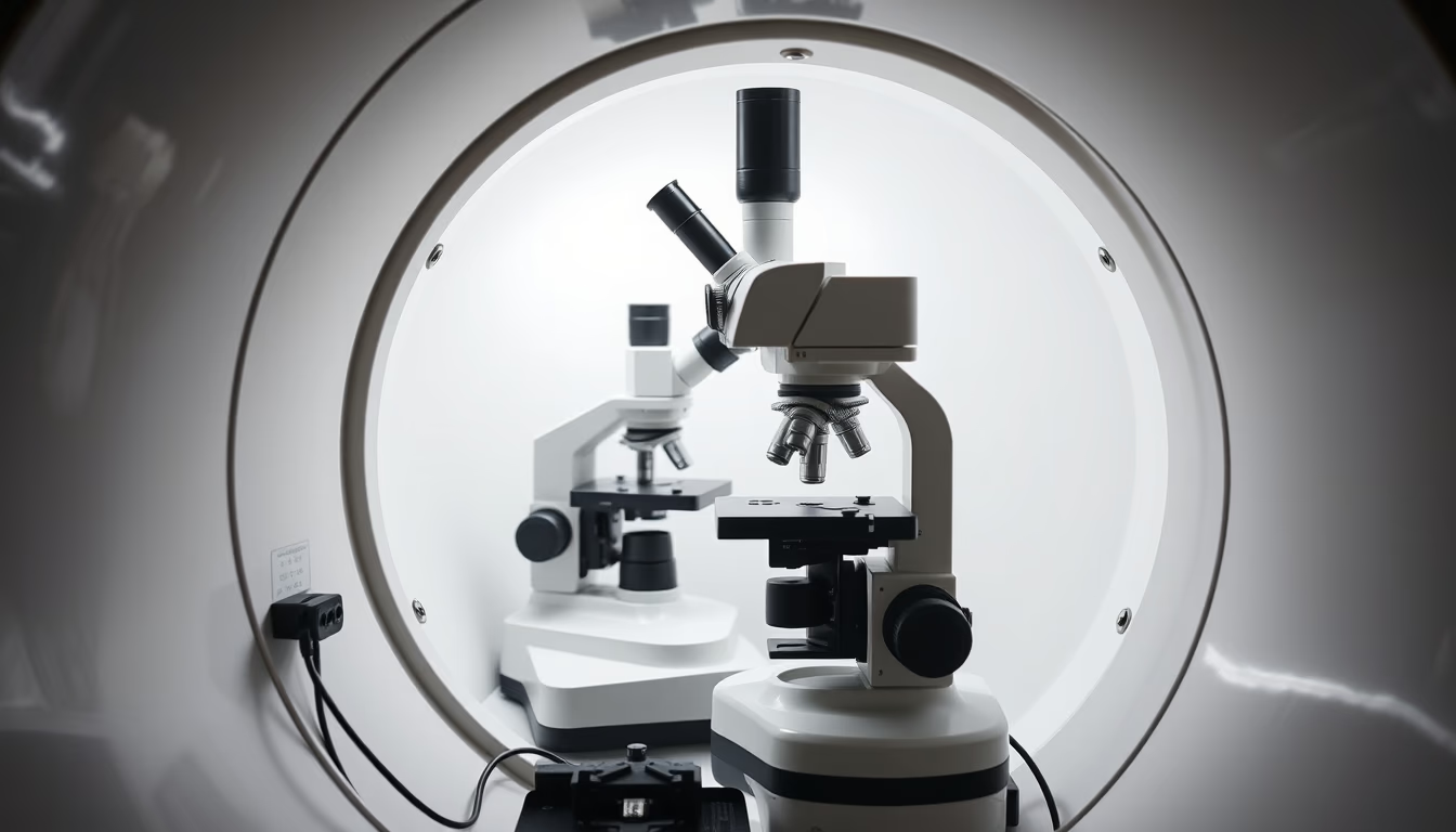
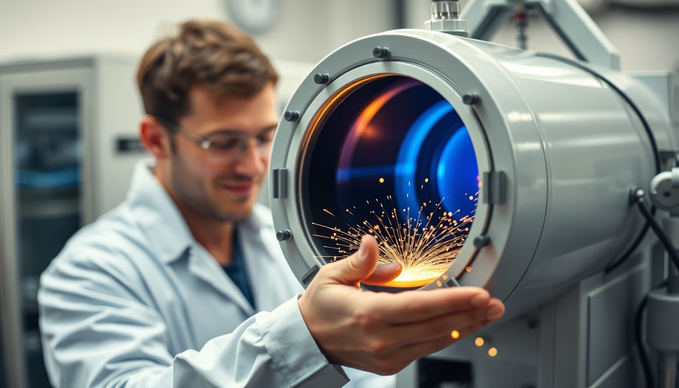
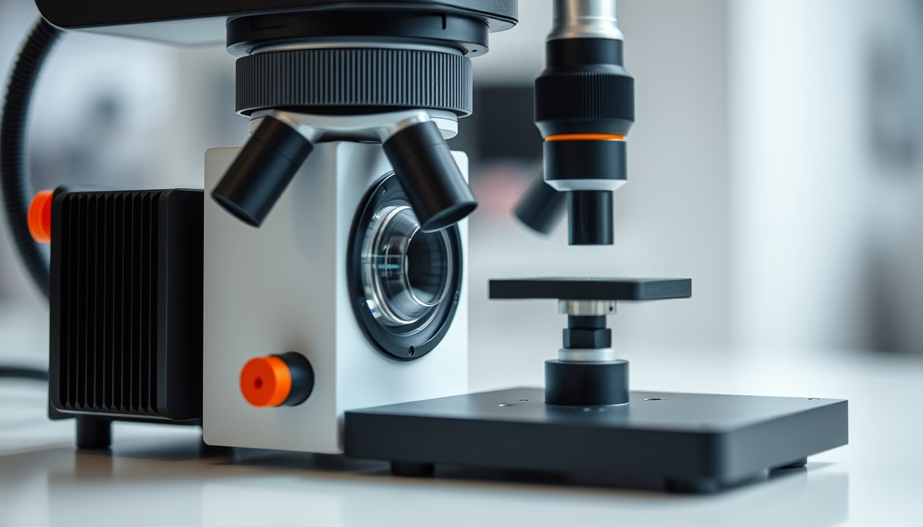
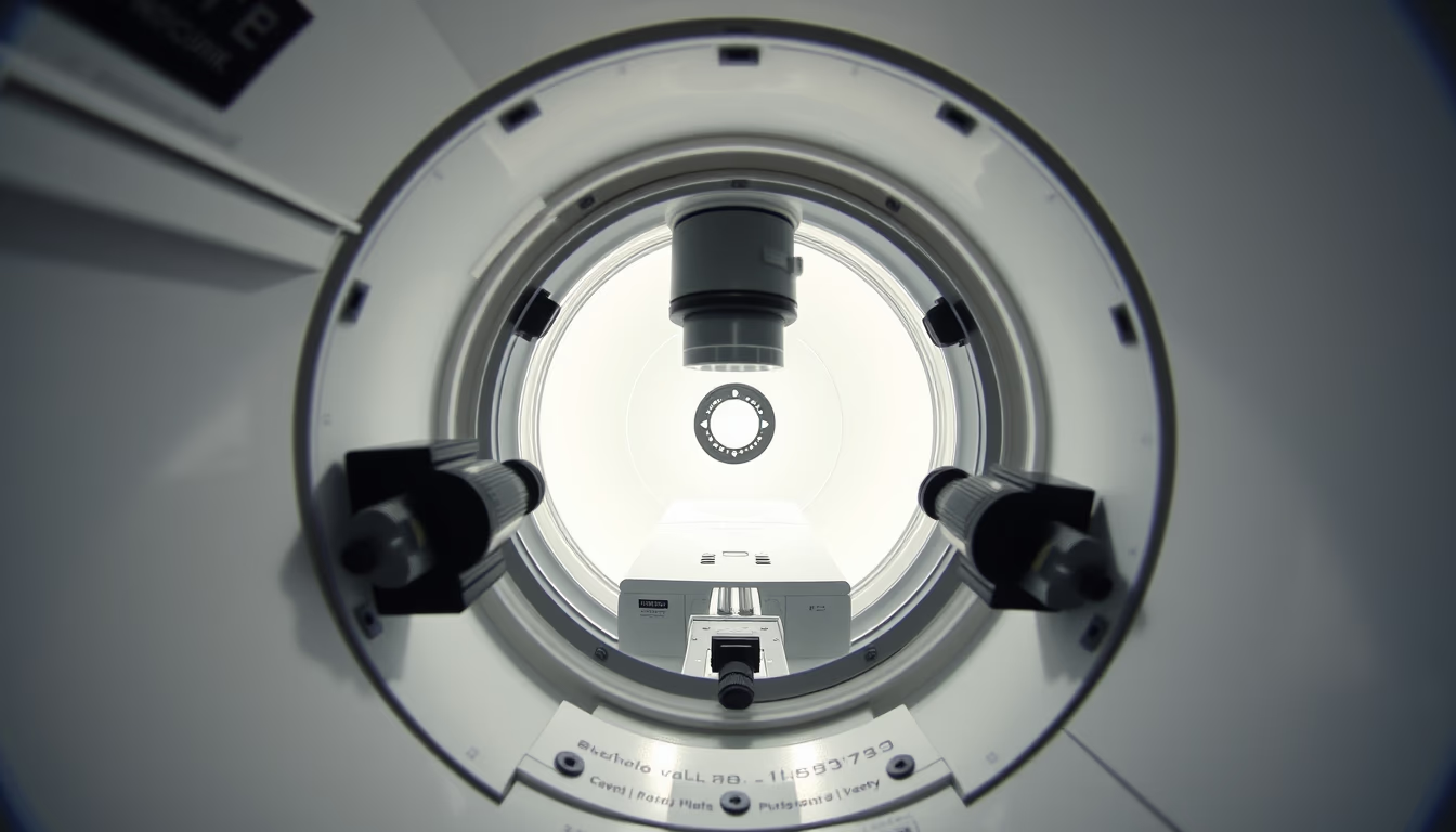
The chamber of an electron microscope is where the sample is analyzed under vacuum, using electron beams for magnifications up to millions of times.
About this subject
The chamber of an electron microscope is a critical component that houses the sample during analysis. Unlike optical microscopes that use light, electron microscopes utilize electron beams to achieve much higher resolutions, capable of visualizing structures at the atomic scale. The chamber must be maintained under vacuum to prevent air molecules from interfering with the electron path, ensuring clear and distortion-free images.
There are two main types of electron microscopes: scanning electron microscopes (SEM) and transmission electron microscopes (TEM). In SEM, the chamber is designed to allow the beam to scan the sample surface, producing high-definition three-dimensional images. In TEM, the chamber is more complex, as the beam passes through a thin sample, requiring an electromagnetic lens system to focus the image. In both cases, the chamber is sealed and equipped with ports for sample insertion, sensors, and detectors.
Temperature and pressure inside the chamber are strictly controlled. Many modern models have cooling systems using liquid nitrogen to reduce thermal vibrations and improve stability. Additionally, the chamber is often coated with materials that absorb scattered electrons, minimizing background noise. Chamber maintenance is essential: any contamination can compromise image quality and damage sensitive components such as the filament and lenses.
Notably, electron microscope chambers are designed to withstand extreme pressure differences. While the interior operates under high vacuum (up to 10^-6 Pa), the exterior is at atmospheric pressure. This requires robust stainless steel walls and special sealing gaskets. Recent advances include chambers with differential vacuum systems, allowing analysis of wet or biological samples without complete dehydration, expanding applications in materials science and biology.
Frequently Asked Questions
Why does the chamber of an electron microscope need to be under vacuum?
The vacuum prevents air molecules from scattering or absorbing electrons, which would degrade image quality. It also protects the filament (electron source) from oxidation and allows proper operation of detectors.
What is the difference between SEM and TEM chambers?
In SEM (scanning electron microscope), the chamber is optimized for the beam to scan the sample surface, often with more space for larger samples. In TEM (transmission electron microscope), the chamber is more complex, with a lens system below the sample and a need for even higher vacuum since the beam passes through the sample.
What materials are used in chamber construction?
The chamber is typically made of stainless steel, resistant to corrosion and able to withstand pressure differences. Internally, it may have coatings like gold or carbon to absorb scattered electrons. Seals are made of special elastomers or metal (for high vacuum).
Direct URL
https://pub-c7d6a6ea828543ac903a74a341ccb2e1.r2.dev/imagens/electron-microscope-chamber-studio-editorial-portrait-fresh2.avifHow to credit
Include a visible link back to UtilizAí. Copy one of the snippets below:
<a href="https://xn--utiliza-eza.com/en/midia/imagens/electron-microscope-chamber-studio-editorial-portrait-fresh2">Electron microscope chamber</a> by <a href="https://xn--utiliza-eza.com">UtilizAí</a>, licensed under <a href="https://creativecommons.org/licenses/by/4.0/">CC BY 4.0</a>.
[Electron microscope chamber](https://xn--utiliza-eza.com/en/midia/imagens/electron-microscope-chamber-studio-editorial-portrait-fresh2) by [UtilizAí](https://xn--utiliza-eza.com), CC BY 4.0
License: CC-BY-4.0