Electron microscope chamber
1344×768 · AVIF · CC BY 4.0
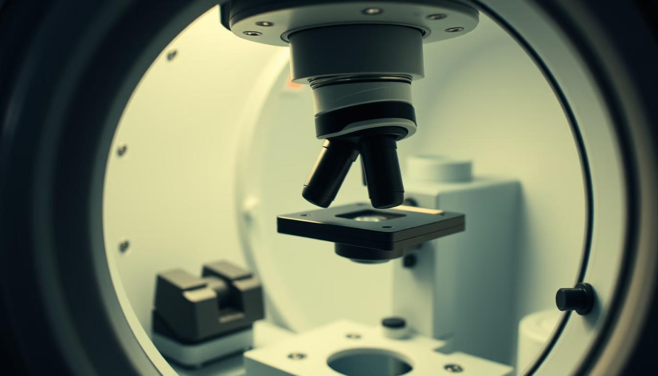
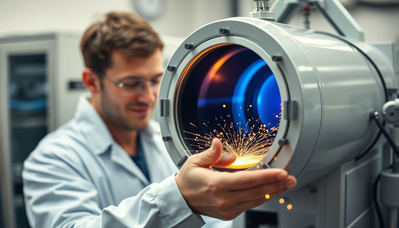
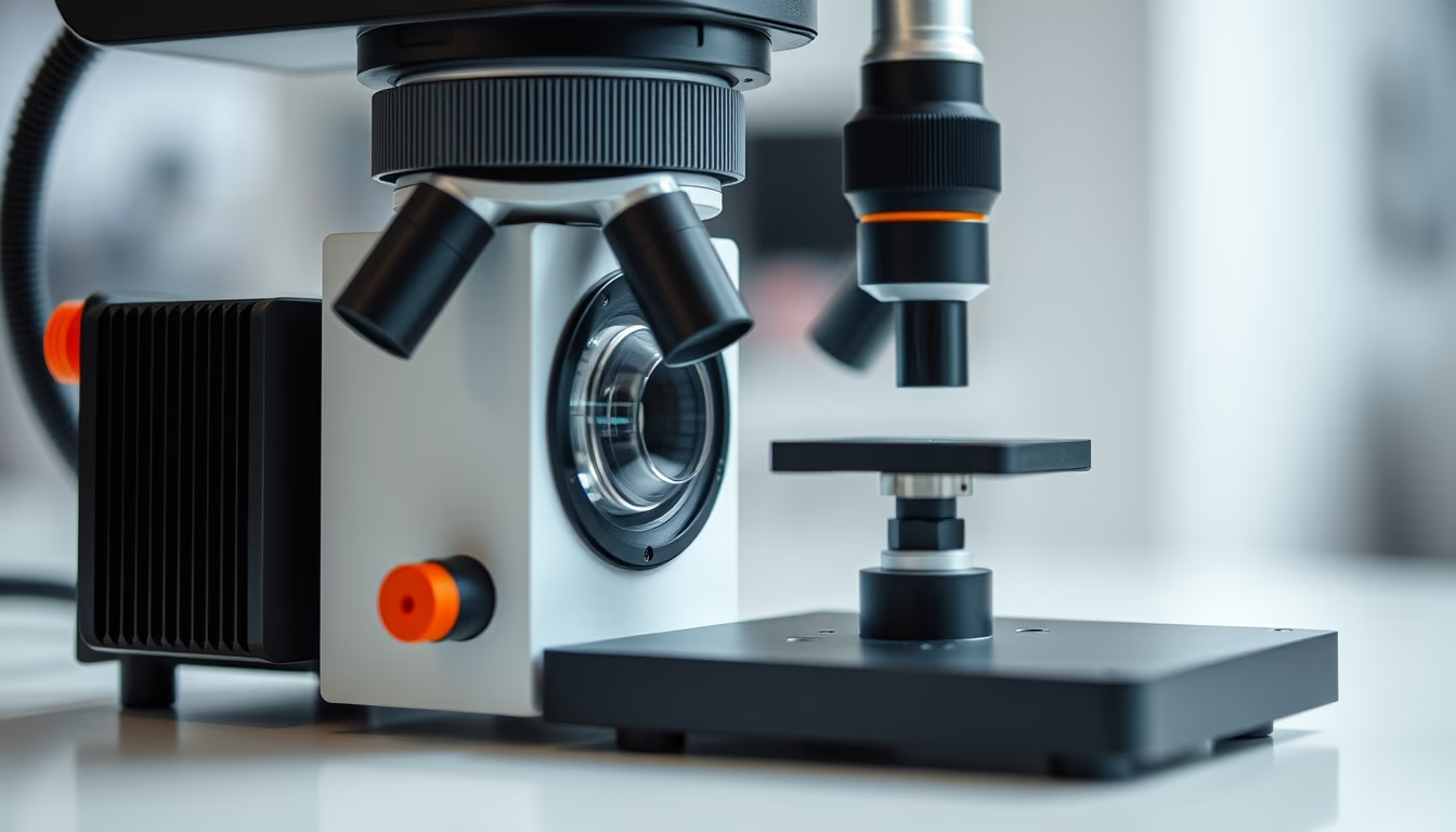
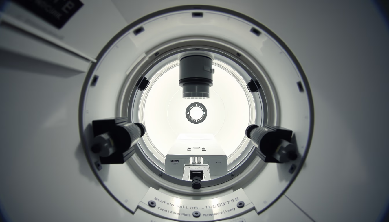
The electron microscope chamber is the vacuum-sealed compartment where the sample is bombarded by electron beams, enabling magnifications up to millions of times.
About this subject
The electron microscope revolutionized materials science and biology by enabling visualization of structures at the nanometer scale. Unlike optical microscopes, which use light, it employs a beam of accelerated electrons, whose much shorter wavelength provides superior resolution. The microscope chamber, where the sample is placed, is maintained under strict vacuum to prevent air molecules from scattering the electrons. There are two main types: scanning electron microscope (SEM), which maps the sample surface, and transmission electron microscope (TEM), which penetrates a thin sample. Sample preparation is crucial and varies by type: for SEM, the sample must be conductive and is often coated with gold; for TEM, it must be ultrathin (less than 100 nm). The first electron microscope was built by Ernst Ruska and Max Knoll in 1931, in Germany, an achievement that earned Ruska the Nobel Prize in Physics in 1986. In Brazil, institutions such as the National Nanotechnology Laboratory (LNNano) in Campinas operate state-of-the-art microscopes for research in nanotechnology, cell biology, and materials science. The chamber, with its pumping systems and detectors, is one of the most critical components, as any contamination or vacuum failure compromises the image.
Frequently Asked Questions
What is the difference between scanning and transmission electron microscopes?
The scanning electron microscope (SEM) scans the sample surface with an electron beam and detects secondary electrons to form a 3D image of the topography. The transmission electron microscope (TEM) lets the beam pass through an ultrathin sample and forms an image from transmitted electrons, revealing internal structures with atomic resolution.
Why does the electron microscope chamber need a vacuum?
Vacuum is necessary because air molecules scatter electrons, reducing resolution and damaging the filament. It also prevents sample contamination and allows electrons to travel from the gun to the detector without interference.
What types of samples can be analyzed with an electron microscope?
Solid materials such as metals, ceramics, polymers, biological tissues, cells, viruses, and even liquid samples in cryomicroscopy can be analyzed. The sample must be stable under vacuum and, for SEM, conductive or coated with a conductive material to avoid charge buildup.
Direct URL
https://pub-c7d6a6ea828543ac903a74a341ccb2e1.r2.dev/imagens/electron-microscope-chamber-lo-fi-disposable-camera-fresh13.avifHow to credit
Include a visible link back to UtilizAí. Copy one of the snippets below:
<a href="https://xn--utiliza-eza.com/en/midia/imagens/electron-microscope-chamber-lo-fi-disposable-camera-fresh13">Electron microscope chamber</a> by <a href="https://xn--utiliza-eza.com">UtilizAí</a>, licensed under <a href="https://creativecommons.org/licenses/by/4.0/">CC BY 4.0</a>.
[Electron microscope chamber](https://xn--utiliza-eza.com/en/midia/imagens/electron-microscope-chamber-lo-fi-disposable-camera-fresh13) by [UtilizAí](https://xn--utiliza-eza.com), CC BY 4.0
License: CC-BY-4.0