Electron microscope chamber
1344×768 · AVIF · CC BY 4.0
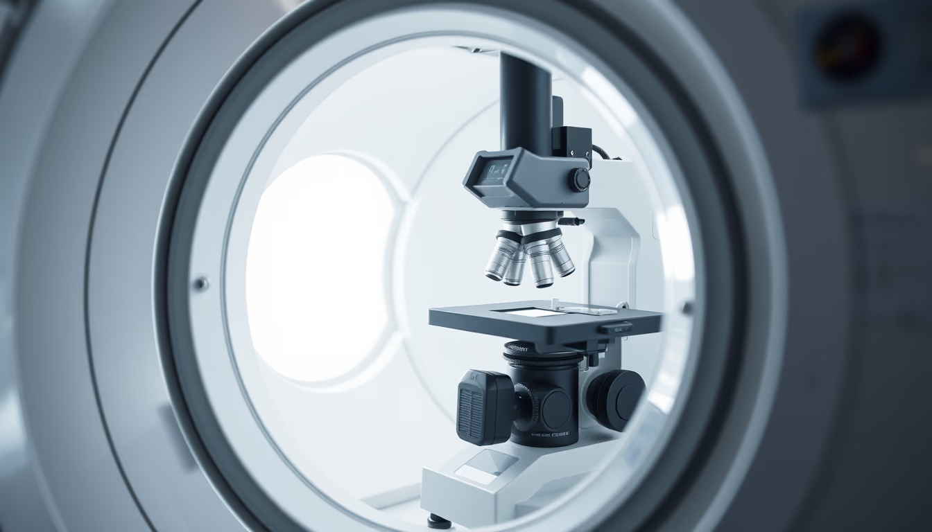
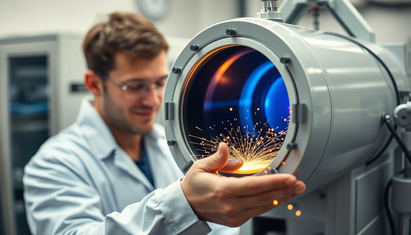
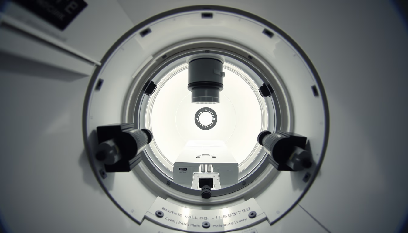
The chamber of an electron microscope is the heart of the equipment, where vacuum allows electron beams to generate images at nanometer scale.
About this subject
The vacuum chamber of an electron microscope is a critical component that houses the sample and detection systems. Unlike optical microscopes that use light, electron microscopes utilize electron beams accelerated by electric and magnetic fields. To ensure that electrons travel a controlled path without colliding with air molecules, the interior must be under high vacuum, typically in the range of 10⁻⁴ to 10⁻⁷ Pa (pascals). This vacuum is maintained by mechanical and turbomolecular pumps, along with sophisticated sealing systems.
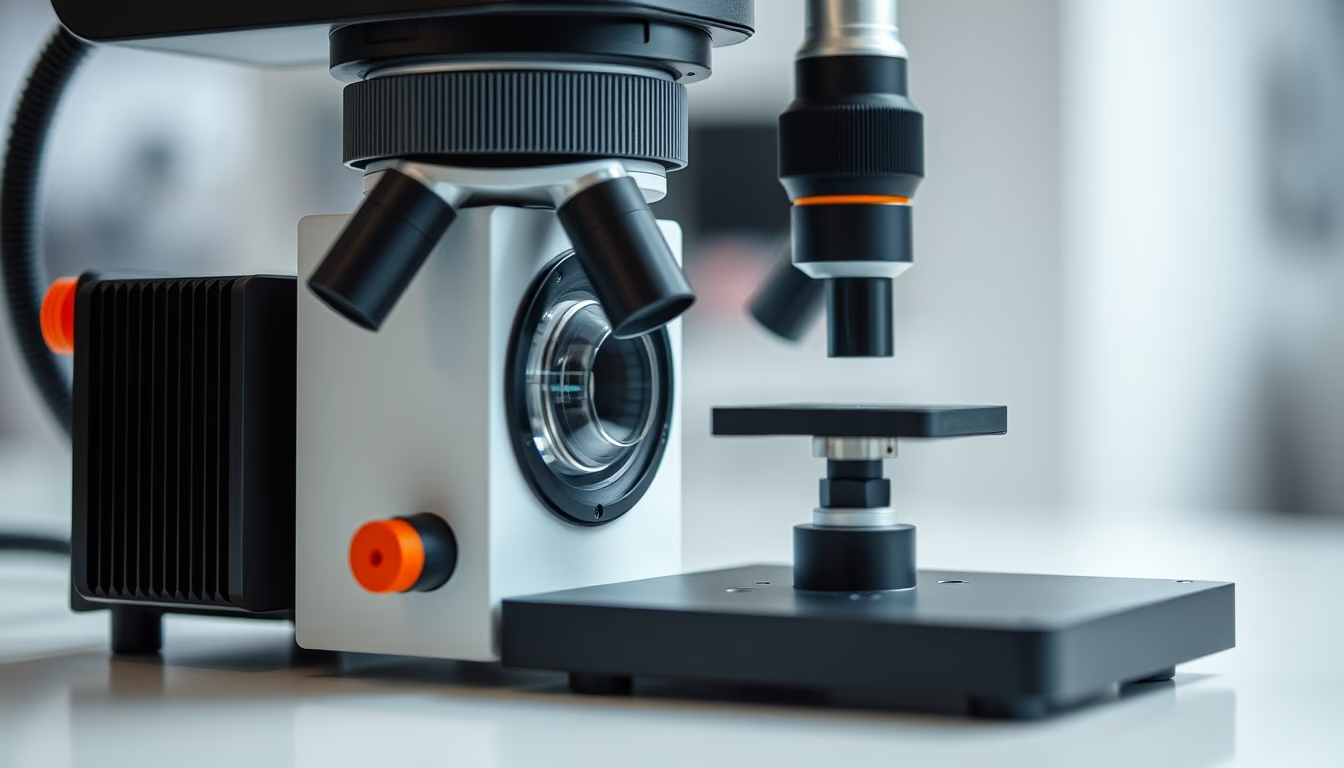
Inside the chamber, the sample is placed on a specimen stage that can be moved with nanometer precision using piezoelectric motors. Electrons interact with the sample, generating signals such as secondary electrons, backscattered electrons, and characteristic X-rays, which are captured by specific detectors. In scanning electron microscopes (SEM), the beam scans the sample surface, while in transmission electron microscopes (TEM), electrons pass through a thin sample. The chamber walls are coated with materials that minimize reflections and electromagnetic interference.
Historically, the development of the vacuum chamber was one of the major technical challenges in building the first electron microscope by Ernst Ruska and Max Knoll in the 1930s. Ruska received the Nobel Prize in Physics in 1986 for this work. Today, modern chambers incorporate cryogenics for sensitive samples, controlled gases for in situ experiments, and even environmental SEMs (ESEM) that operate without full vacuum. Precision control of pressure and temperature within the chamber determines the final image quality, with resolutions reaching 0.05 nm.
Interestingly, some electron microscopes have fast-loading chambers that allow sample exchange without breaking the main vacuum, optimizing workflow. The chamber also houses beam alignment systems, such as magnetic lenses and apertures, which focus the electrons. In materials science, biology, and nanotechnology labs, the electron microscope chamber is where cutting-edge research happens, revealing structures previously invisible.
Frequently Asked Questions
What is the typical pressure inside an electron microscope chamber?
Pressure ranges from 10⁻⁴ to 10⁻⁷ Pa (pascals), depending on the microscope type. Transmission electron microscopes (TEM) require a more stringent vacuum of about 10⁻⁶ to 10⁻⁷ Pa, while scanning electron microscopes (SEM) operate at slightly higher pressures, between 10⁻⁴ and 10⁻⁶ Pa.
Why is vacuum necessary in an electron microscope?
Vacuum prevents air molecules from scattering or deflecting the electron beam, ensuring it reaches the sample in a controlled manner. It also protects electronic components and prevents sample contamination.
What types of detectors are found inside the chamber?
Key detectors include secondary electron (SE) and backscattered electron (BSE) detectors in SEM, and X-ray detectors for elemental analysis (EDS). In TEM, there are transmitted electron detectors and CCD or CMOS cameras for imaging.
Direct URL
https://pub-c7d6a6ea828543ac903a74a341ccb2e1.r2.dev/imagens/electron-microscope-chamber-documentary-photography-fresh0.avifHow to credit
Include a visible link back to UtilizAí. Copy one of the snippets below:
<a href="https://xn--utiliza-eza.com/en/midia/imagens/electron-microscope-chamber-documentary-photography-fresh0">Electron microscope chamber</a> by <a href="https://xn--utiliza-eza.com">UtilizAí</a>, licensed under <a href="https://creativecommons.org/licenses/by/4.0/">CC BY 4.0</a>.
[Electron microscope chamber](https://xn--utiliza-eza.com/en/midia/imagens/electron-microscope-chamber-documentary-photography-fresh0) by [UtilizAí](https://xn--utiliza-eza.com), CC BY 4.0
License: CC-BY-4.0