Electron microscope chamber
1344×768 · AVIF · CC BY 4.0
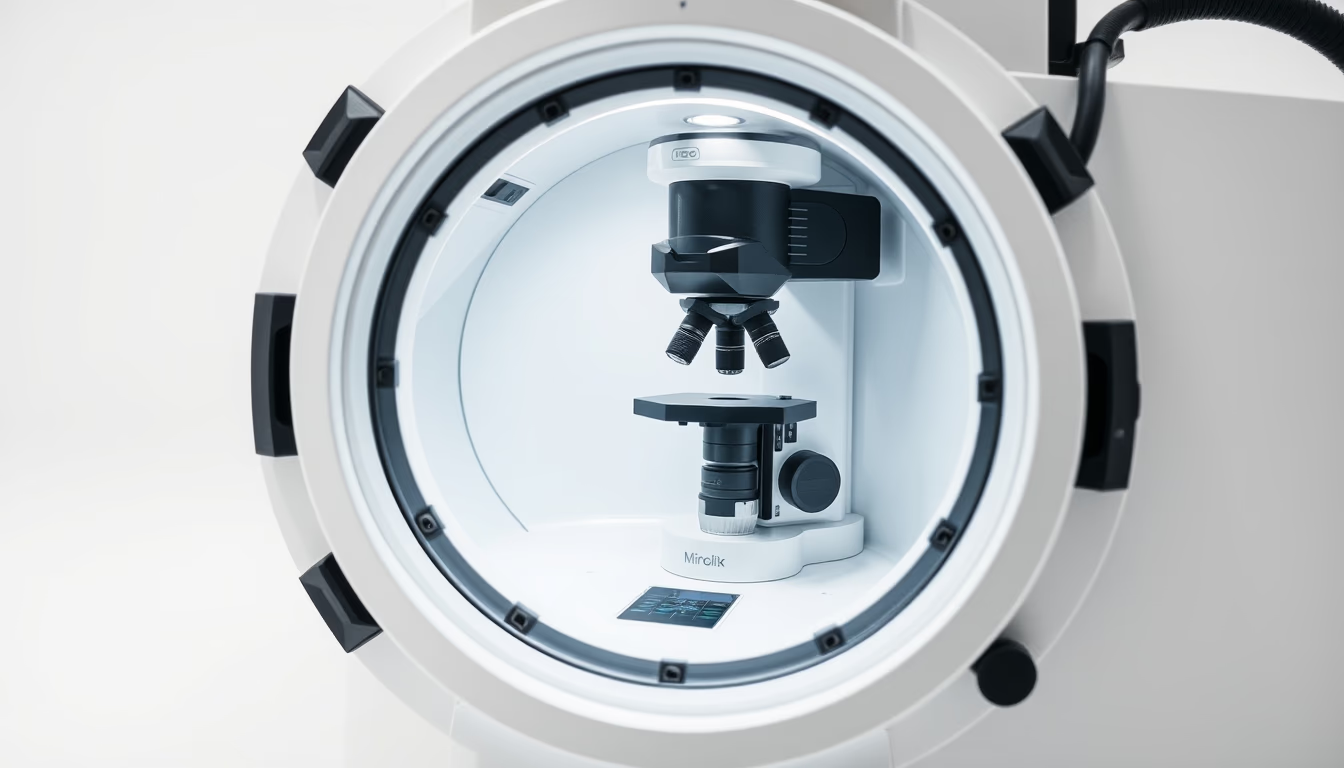
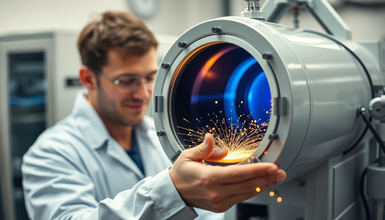
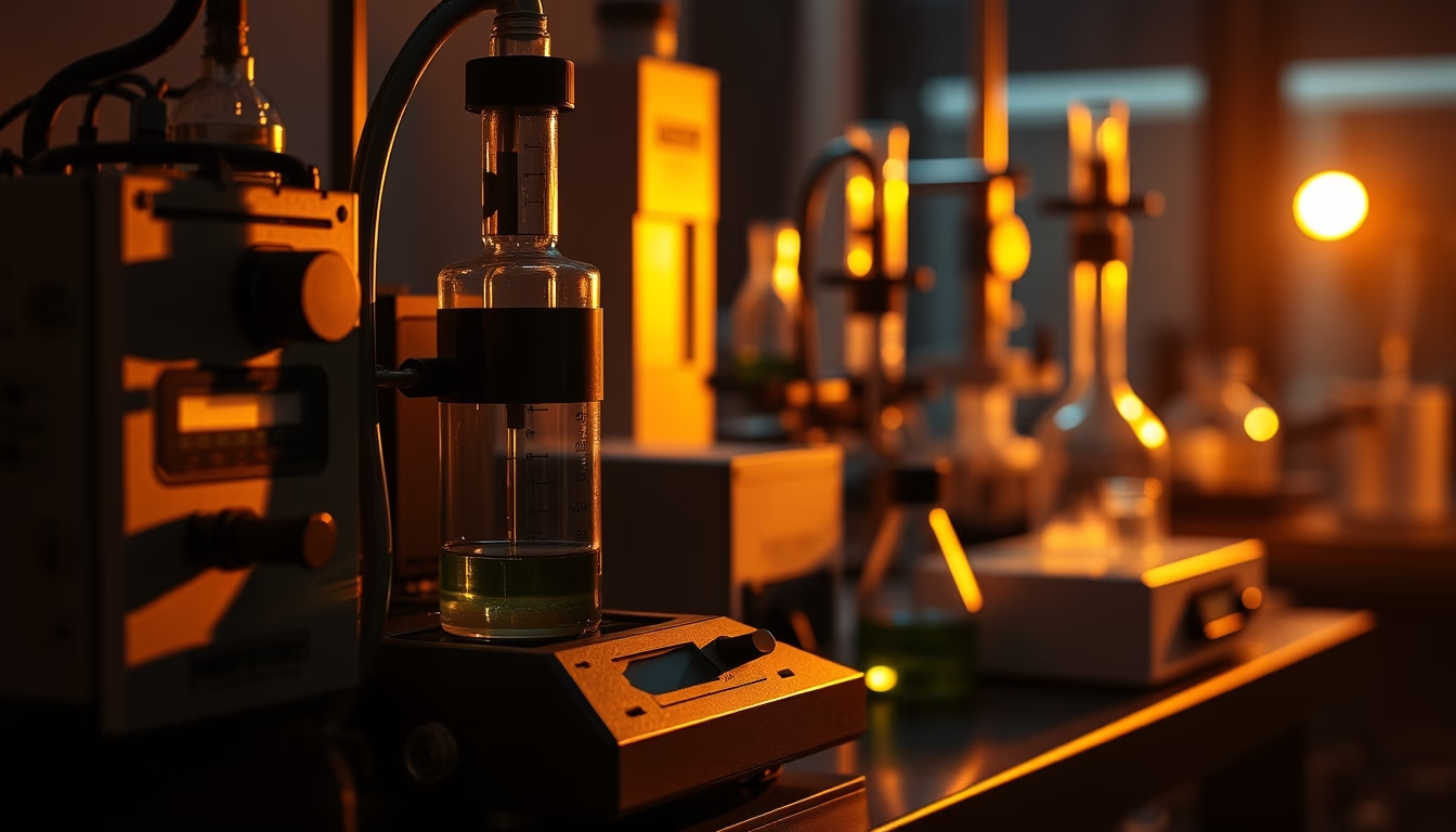
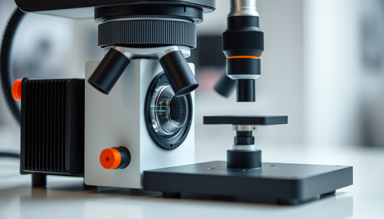
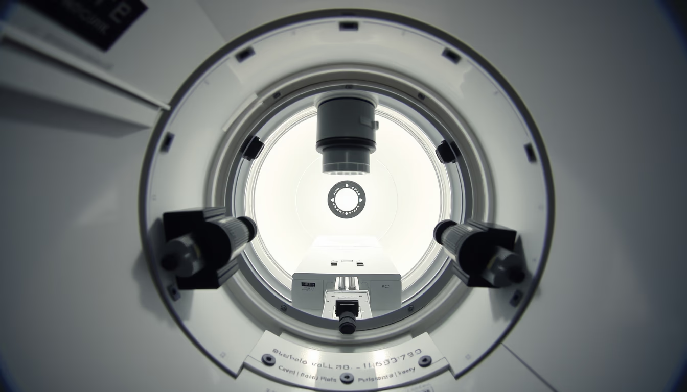
The electron microscope chamber is a sealed vacuum environment where the sample is placed for observation by an electron beam, crucial to avoid interference.
About this subject
The electron microscope chamber is a core component in scanning electron microscopes (SEM) and transmission electron microscopes (TEM). Its primary role is to maintain a high vacuum, typically between 10⁻⁴ and 10⁻⁷ Pa, allowing the electron beam to travel without colliding with air molecules, which would degrade image quality and damage the instrument. The chamber is made of stainless steel to withstand external atmospheric pressure and resist corrosion. It features glass or transparent windows for visual inspection, along with ports for attaching detectors, manipulators, and cryogenic systems.
Sample introduction is performed via a transfer chamber, which pumps out air before opening the main chamber, preserving the vacuum. Inside, a motorized stage positions the sample with nanometer precision. For biological specimens, a cryogenic stage cooled with liquid nitrogen is often used to maintain hydration and prevent damage from dehydration. The chamber also houses detectors for secondary or backscattered electrons, capturing signals generated by the beam's interaction with the sample.
The evolution of electron microscope chambers has paralleled the development of electron microscopy since the 1930s, with advances in sealing, vacuum pumps, and materials. Modern turbomolecular vacuum systems achieve ultra-low pressures in seconds, and special coatings reduce contaminant adsorption. The chamber is a key factor determining image resolution and stability, designed to minimize vibrations and stray electric fields.
Frequently Asked Questions
Why is vacuum necessary in the electron microscope chamber?
Vacuum prevents air molecules from interacting with the electron beam, which would cause scattering and loss of resolution. It also protects the filament and reduces sample contamination.
How is the sample inserted into the chamber without breaking the vacuum?
A transfer chamber or airlock is used. The sample is placed in this compartment, which is pumped separately. Once vacuum is reached, a valve opens and the sample is moved into the main chamber.
What materials make up the chamber and why?
The chamber is typically made of stainless steel for mechanical strength and low outgassing. Copper or elastomer seals are used for gaskets, and windows are borosilicate glass or sapphire for transparency and airtightness.
Direct URL
https://pub-c7d6a6ea828543ac903a74a341ccb2e1.r2.dev/imagens/electron-microscope-chamber-commercial-product-photography-fresh8.avifHow to credit
Include a visible link back to UtilizAí. Copy one of the snippets below:
<a href="https://xn--utiliza-eza.com/en/midia/imagens/electron-microscope-chamber-commercial-product-photography-fresh8">Electron microscope chamber</a> by <a href="https://xn--utiliza-eza.com">UtilizAí</a>, licensed under <a href="https://creativecommons.org/licenses/by/4.0/">CC BY 4.0</a>.
[Electron microscope chamber](https://xn--utiliza-eza.com/en/midia/imagens/electron-microscope-chamber-commercial-product-photography-fresh8) by [UtilizAí](https://xn--utiliza-eza.com), CC BY 4.0
License: CC-BY-4.0