Electron microscope chamber
1344×768 · AVIF · CC BY 4.0
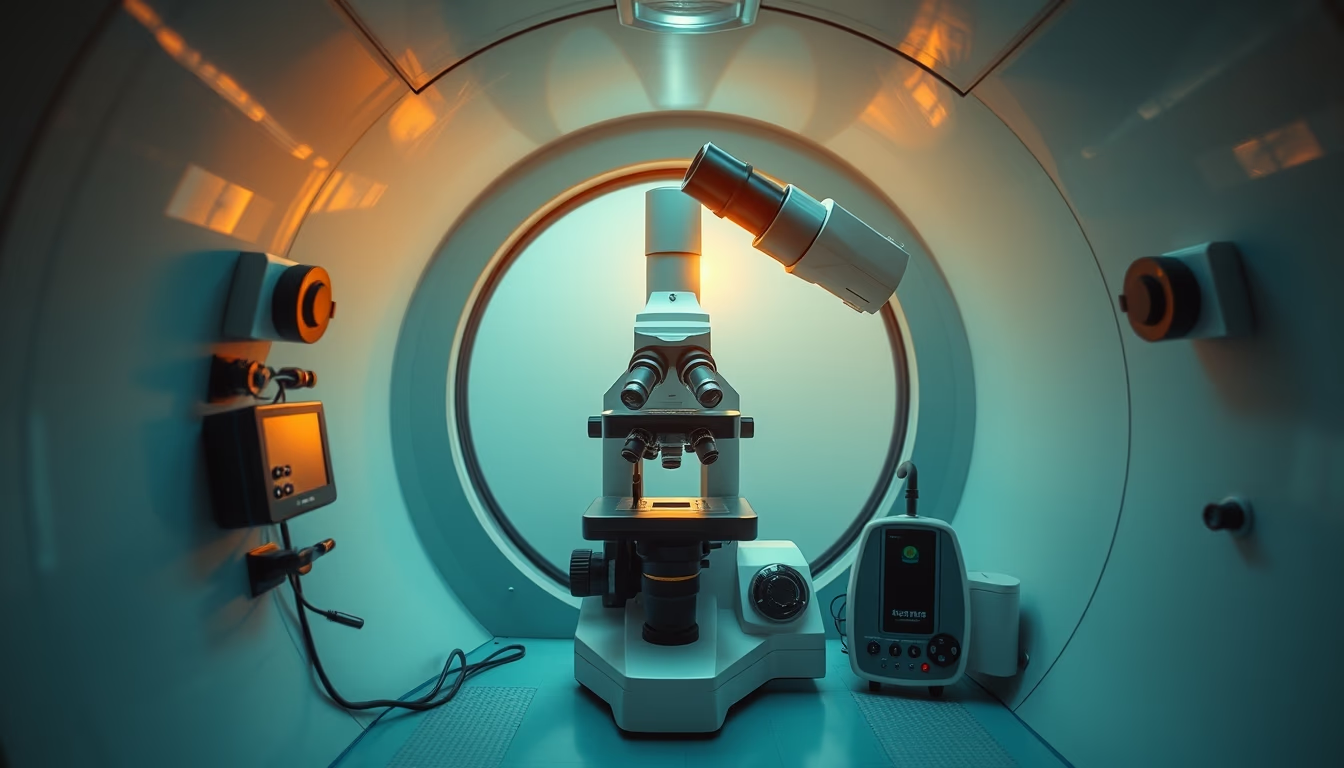
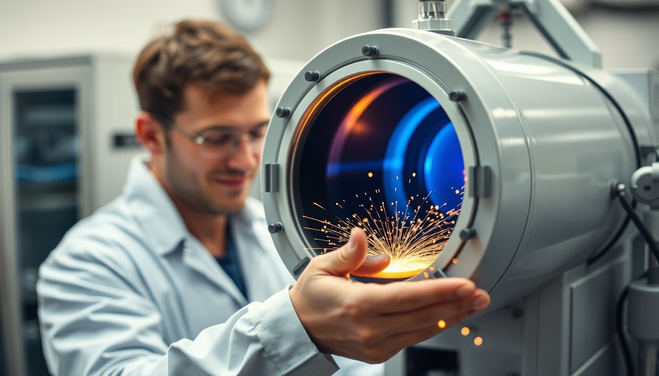
The electron microscope chamber is the heart of the instrument, where the electron beam interacts with the sample under ultra-high vacuum, enabling magnifications up to 50 million times.
About this subject
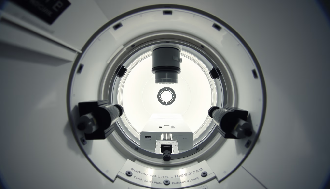
The electron microscope chamber is a sealed compartment designed to maintain ultra-high vacuum conditions, essential for the electron beam to function properly. Unlike optical microscopes that use light, electron microscopes use a beam of electrons accelerated by electric fields. The chamber houses the sample and the electromagnetic lenses that focus the beam, ensuring that electrons do not collide with air molecules, which would scatter the beam and degrade the image. This vacuum, ranging from 10^-4 to 10^-9 Pascals, is maintained by mechanical and diffusion or turbomolecular pumps.
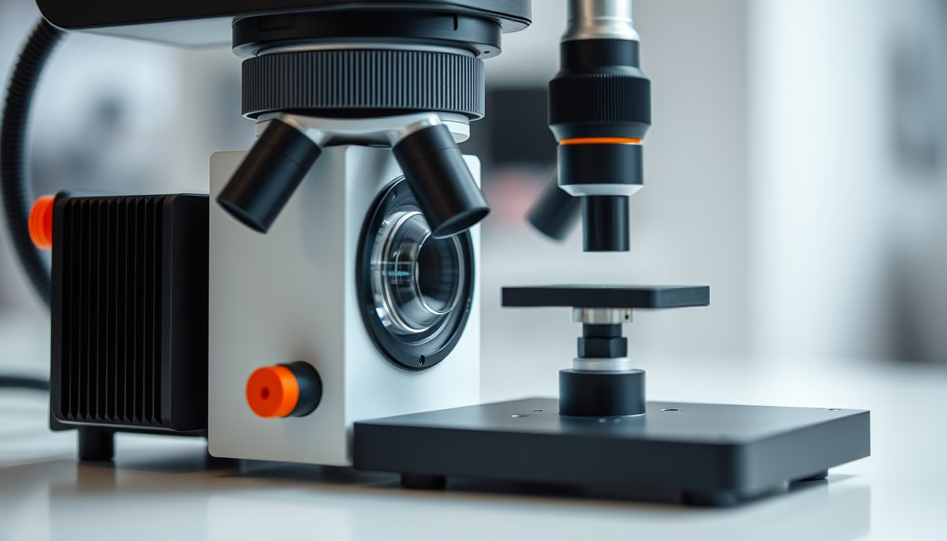
Inside the chamber, the sample is placed on a movable stage that allows rotation and tilting, enabling three-dimensional visualization. Depending on the microscope type, the chamber may contain detectors for secondary or backscattered electrons, as well as X-ray spectrometers for elemental analysis. In transmission electron microscopes (TEM), the chamber is elongated, with the beam passing through an ultrathin sample before hitting a fluorescent screen or detector. In scanning electron microscopes (SEM), the chamber is wider to accommodate larger samples.
The chamber also plays a key role in thermal and mechanical stability. Temperature variations can cause thermal drift, shifting the sample sub-micrometrically. Therefore, many chambers are cooled and mounted on vibration-damping tables. Applications include analysis of semiconductor materials, nanoparticles, biological tissues, and fossils. Recently, chambers with controlled gas environments (environmental microscopy) allow observation of wet samples or chemical reactions in real time, expanding potential for studies in catalysis and cell biology.
Frequently Asked Questions
What is the difference between the chamber of a scanning electron microscope and a transmission electron microscope?
In SEM, the chamber is more spacious to accommodate larger samples and the beam scans the surface. In TEM, the chamber is narrower and elongated, as the beam passes through an ultrathin sample. The vacuum and detector systems also differ based on the type of electron-sample interaction.
Why is vacuum essential in the electron microscope chamber?
Vacuum prevents air molecules from deflecting or absorbing the electron beam, ensuring a free path and sharp image. It also protects the tungsten filament or electron gun from oxidation and allows high acceleration voltages without electrical discharge.
What are the current magnification limits of electron microscopes?
Aberration-corrected transmission electron microscopes can achieve magnifications up to 50 million times with sub-angstrom resolution (below 0.1 nm). Scanning electron microscopes reach about 2 million times with nanometric resolution.
Direct URL
https://pub-c7d6a6ea828543ac903a74a341ccb2e1.r2.dev/imagens/electron-microscope-chamber-cinematic-wide-shot-fresh1.avifHow to credit
Include a visible link back to UtilizAí. Copy one of the snippets below:
<a href="https://xn--utiliza-eza.com/en/midia/imagens/electron-microscope-chamber-cinematic-wide-shot-fresh1">Electron microscope chamber</a> by <a href="https://xn--utiliza-eza.com">UtilizAí</a>, licensed under <a href="https://creativecommons.org/licenses/by/4.0/">CC BY 4.0</a>.
[Electron microscope chamber](https://xn--utiliza-eza.com/en/midia/imagens/electron-microscope-chamber-cinematic-wide-shot-fresh1) by [UtilizAí](https://xn--utiliza-eza.com), CC BY 4.0
License: CC-BY-4.0