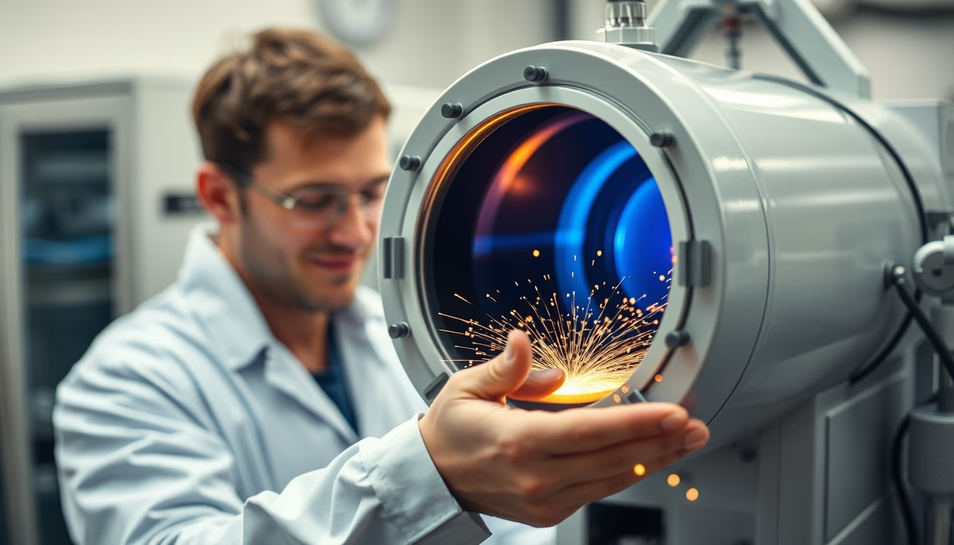
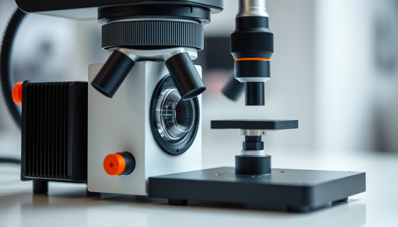
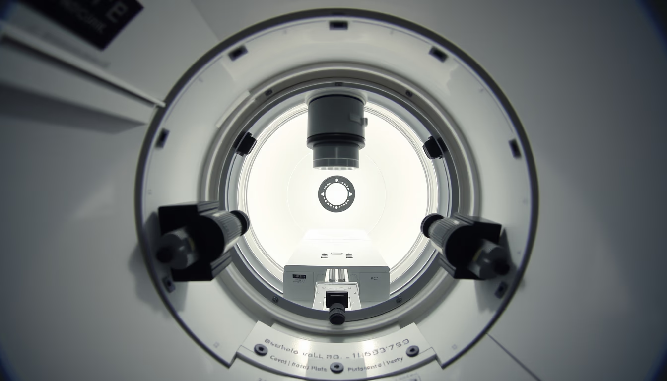
Electron microscope chamber
1344×768 · AVIF · CC BY 4.0
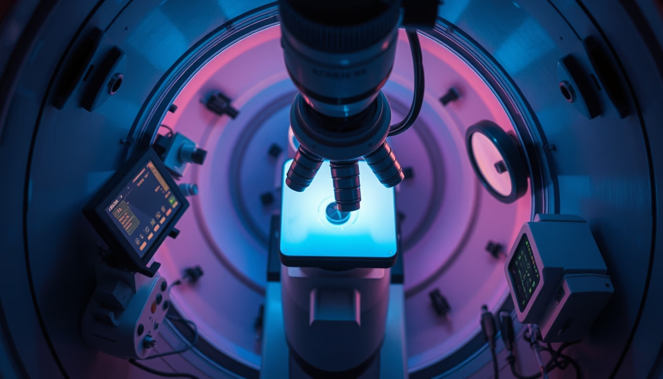
The electron microscope chamber is a vacuum-sealed component that houses the sample during analysis by electron beam in scanning or transmission electron microscopy.
About this subject
The electron microscope chamber is the core of instruments like the scanning electron microscope (SEM) and transmission electron microscope (TEM). It is a hermetically sealed compartment where the sample is placed and maintained under high vacuum, a condition essential for the electron beam to interact with the material without being scattered by air molecules. Without this vacuum, high resolution images reaching the nanometer scale would be impossible. The interior houses precision motorized stages, secondary and backscattered electron detectors, and cryogenic systems for heat sensitive specimens.
Historically, the development of the vacuum chamber paralleled the evolution of electron microscopes themselves. The first functional model was built by Ernst Ruska and Max Knoll in 1931 in Germany, but modern chambers employ turbomolecular pumps and metal seals to achieve pressures around 10⁻⁴ Pa. In environmental microscopy, the chamber can operate with controlled gas pressures such as water vapor, enabling observation of hydrated materials or in situ reactions. This is crucial for fields like cell biology and catalysis.
Technically and culturally, the electron microscope chamber symbolizes the advancement of materials science and nanotechnology. Institutions like the Brazilian Nanotechnology National Laboratory (LNNano) use these chambers for research ranging from fractography to semiconductor development. A little known fact is that some recent models integrate focused ion beam (FIB) systems inside the same chamber, allowing in situ cutting and modification of samples while observing.
The versatility of these chambers is also seen in accessories such as sample holders for heating, cooling, or applying electric fields. Design must minimize vibrations and stray magnetic fields to ensure stability for magnifications exceeding 1,000,000 times. Dust and contamination are removed by plasma cleaning, and maintenance requires periodic gasket replacement. Undoubtedly, the chamber is the element that makes electron microscopy an indispensable tool in modern science.
Frequently Asked Questions
Why does the electron microscope chamber need a vacuum?
The vacuum prevents air molecules from deflecting or scattering the electron beam, ensuring it reaches the sample focused and produces high resolution images, while also protecting the electron source filament from oxidation.
What types of samples can be analyzed inside the chamber?
Conductive materials are ideal, but non conductive samples can be coated with gold or carbon. Biological specimens must be dehydrated and fixed. Environmental chambers allow hydrated materials or reactions in controlled gases.
What is the typical pressure inside an electron microscope chamber?
For conventional microscopy, pressure ranges from 10⁻³ to 10⁻⁴ Pa. High vacuum systems reach 10⁻⁶ Pa. Environmental microscopy operates at higher pressures, up to a few hundred Pascals.
Direct URL
https://pub-c7d6a6ea828543ac903a74a341ccb2e1.r2.dev/imagens/electron-microscope-chamber-aerial-drone-shot-fresh3.avifHow to credit
Include a visible link back to UtilizAí. Copy one of the snippets below:
<a href="https://xn--utiliza-eza.com/en/midia/imagens/electron-microscope-chamber-aerial-drone-shot-fresh3">Electron microscope chamber</a> by <a href="https://xn--utiliza-eza.com">UtilizAí</a>, licensed under <a href="https://creativecommons.org/licenses/by/4.0/">CC BY 4.0</a>.
[Electron microscope chamber](https://xn--utiliza-eza.com/en/midia/imagens/electron-microscope-chamber-aerial-drone-shot-fresh3) by [UtilizAí](https://xn--utiliza-eza.com), CC BY 4.0
License: CC-BY-4.0