Nucleus chromosomes microscope
1344×768 · AVIF · CC BY 4.0
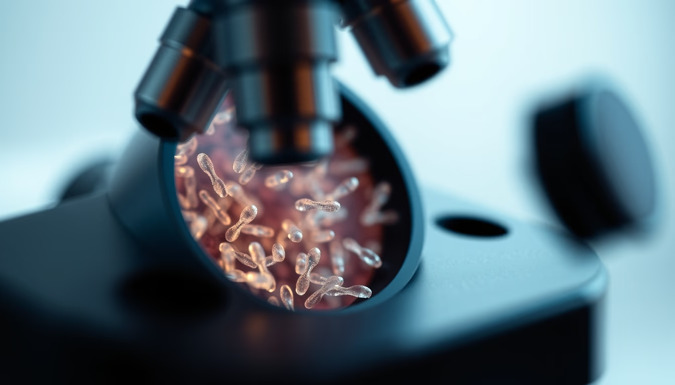
The cell nucleus houses chromosomes, structures that carry DNA. Understand their organization and function within the cell.
About this subject
The nucleus is an essential organelle in eukaryotic cells, acting as a control center that stores genetic material. Inside it, chromosomes are organized into a complex structure of DNA and histone proteins, forming chromatin. During cell division, chromatin condenses into chromosomes visible under a microscope, allowing equal segregation of the genome. In humans, each somatic cell contains 46 chromosomes, arranged in 23 pairs, inherited from each parent.
Light microscopy, especially with specific stains like Giemsa, reveals characteristic bands on chromosomes, used to identify chromosomal abnormalities such as Down syndrome (trisomy 21). More advanced techniques, such as fluorescence microscopy with DNA probes, allow visualization of specific genes and structural rearrangements. Observing chromosomes in metaphase, when they are most condensed, is fundamental for karyotyping, a test that analyzes chromosome number and morphology.
Chromosomes also play a crucial role in heredity. Each gene occupies a specific locus on a chromosome, and recombination during meiosis generates genetic variability. The discovery of the DNA double helix structure by Watson and Crick in 1953 revolutionized understanding of how chromosomes store and transmit information. Today, sequencing and gene editing techniques like CRISPR allow direct manipulation of chromosomal DNA, opening therapeutic possibilities.
Interestingly, not all chromosomes are the same across species. While the fruit fly has only 8 chromosomes, some plants like wheat have 42. Nuclear organization also varies: in specialized cells like neurons, the position of chromosomes within the nucleus can influence gene expression. Ongoing study of chromosome structure and function is vital for genetic medicine and biotechnology.
Frequently Asked Questions
What are chromosomes?
Chromosomes are structures made of DNA and proteins that contain genes, located in the nucleus of eukaryotic cells. They become visible during cell division.
How many chromosomes do humans have?
Humans have 46 chromosomes in each somatic cell, arranged in 23 pairs. One pair consists of the sex chromosomes (XX or XY).
How are chromosomes visualized under a microscope?
Chromosomes are stained with substances like Giemsa and observed under a light microscope during metaphase, when they are most condensed. Fluorescence techniques allow marking specific genes.
Direct URL
https://pub-c7d6a6ea828543ac903a74a341ccb2e1.r2.dev/imagens/nucleus-chromosomes-microscope-macro-closeup-p1.avifHow to credit
Include a visible link back to UtilizAí. Copy one of the snippets below:
<a href="https://xn--utiliza-eza.com/en/midia/imagens/nucleus-chromosomes-microscope-macro-closeup-p1">Nucleus chromosomes microscope</a> by <a href="https://xn--utiliza-eza.com">UtilizAí</a>, licensed under <a href="https://creativecommons.org/licenses/by/4.0/">CC BY 4.0</a>.
[Nucleus chromosomes microscope](https://xn--utiliza-eza.com/en/midia/imagens/nucleus-chromosomes-microscope-macro-closeup-p1) by [UtilizAí](https://xn--utiliza-eza.com), CC BY 4.0
License: CC-BY-4.0





