Mri scanner machine in radiology department
1344×768 · AVIF · CC BY 4.0
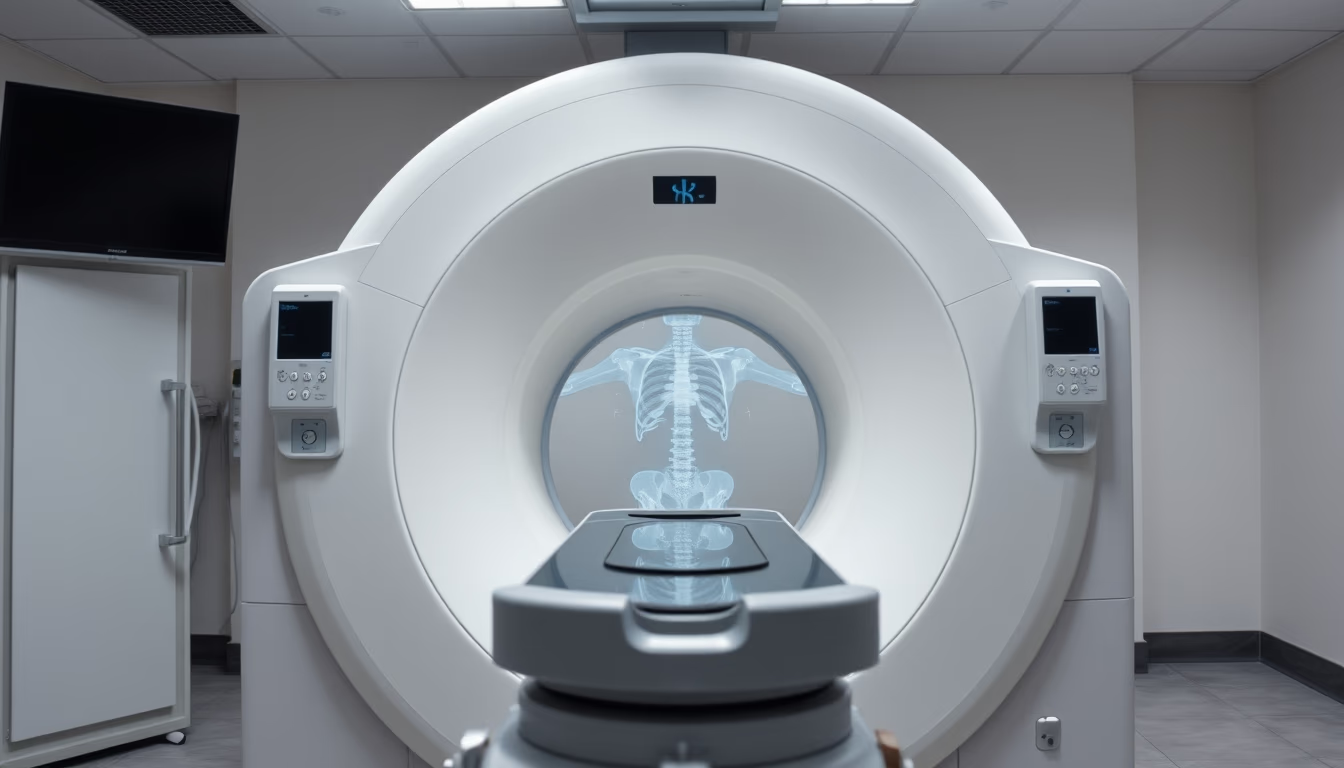
An MRI scanner machine operating in a hospital radiology department, highlighting non-invasive imaging technology.
About this subject
Magnetic resonance imaging (MRI) is a medical imaging technique that uses strong magnetic fields and radio waves to generate detailed images of organs and tissues within the human body. Unlike X-rays and computed tomography, MRI does not use ionizing radiation, making it especially safe for repeated exams and vulnerable populations such as children and pregnant women. The equipment, as depicted in the image, consists of a large cylindrical magnet that creates a uniform magnetic field, typically between 1.5 and 3 teslas in standard hospitals, though 7-tesla machines are used in research.
MRI is essential for diagnosing neurological, musculoskeletal, and oncological conditions, allowing visualization of tumors, spinal cord injuries, joint diseases, and brain anomalies with high soft-tissue contrast. The patient is positioned inside the scanner and must remain still during image acquisition, which can take 15 to 60 minutes depending on the area being examined. One technical challenge is the noise generated by the magnetic field gradient, producing loud tapping or buzzing sounds that require ear protection.
Historically, MRI was developed from the principles of nuclear magnetic resonance, discovered in the 1940s by physicists Felix Bloch and Edward Purcell, who won the Nobel Prize in Physics in 1952. The first whole-body scanner was built in the 1970s, and since then technology has evolved to faster systems with higher resolution. Today, MRI is considered the gold standard for many clinical indications, although its high cost and the need for magnetically shielded environments limit availability in less developed regions.
In Brazil, MRI is available in large hospitals and specialized clinics, mainly in capitals and major urban centers. The public health system (SUS) offers the exam, but waiting lists can be long due to high demand. The image, with professional lighting and symmetric composition, is typical of educational or science communication materials, showing the scanner in use position with the bed extended and the portal open to receive the patient.
Frequently Asked Questions
Is an MRI scan painful?
No, the exam is painless. The patient only needs to lie still inside the scanner. There may be discomfort from staying still for a long time or anxiety from the confined space, but no physical pain.
Who cannot have an MRI scan?
People with non-MRI-compatible metal implants, such as old pacemakers, aneurysm clips, or metal fragments in the eye, cannot undergo the exam. It is essential to inform the doctor about any metal in the body.
How long does an MRI scan take?
The duration ranges from 15 to 60 minutes, depending on the body part and the number of sequences needed. More complex exams, such as abdominal or with contrast, may take longer.
Direct URL
https://pub-c7d6a6ea828543ac903a74a341ccb2e1.r2.dev/imagens/mri-scanner-machine-in-radiology-department-symmetric-composition-studio-professional-lighting.avifHow to credit
Include a visible link back to UtilizAí. Copy one of the snippets below:
<a href="https://xn--utiliza-eza.com/en/midia/imagens/mri-scanner-machine-in-radiology-department-symmetric-composition-studio-professional-lighting">Mri scanner machine in radiology department</a> by <a href="https://xn--utiliza-eza.com">UtilizAí</a>, licensed under <a href="https://creativecommons.org/licenses/by/4.0/">CC BY 4.0</a>.
[Mri scanner machine in radiology department](https://xn--utiliza-eza.com/en/midia/imagens/mri-scanner-machine-in-radiology-department-symmetric-composition-studio-professional-lighting) by [UtilizAí](https://xn--utiliza-eza.com), CC BY 4.0
License: CC-BY-4.0