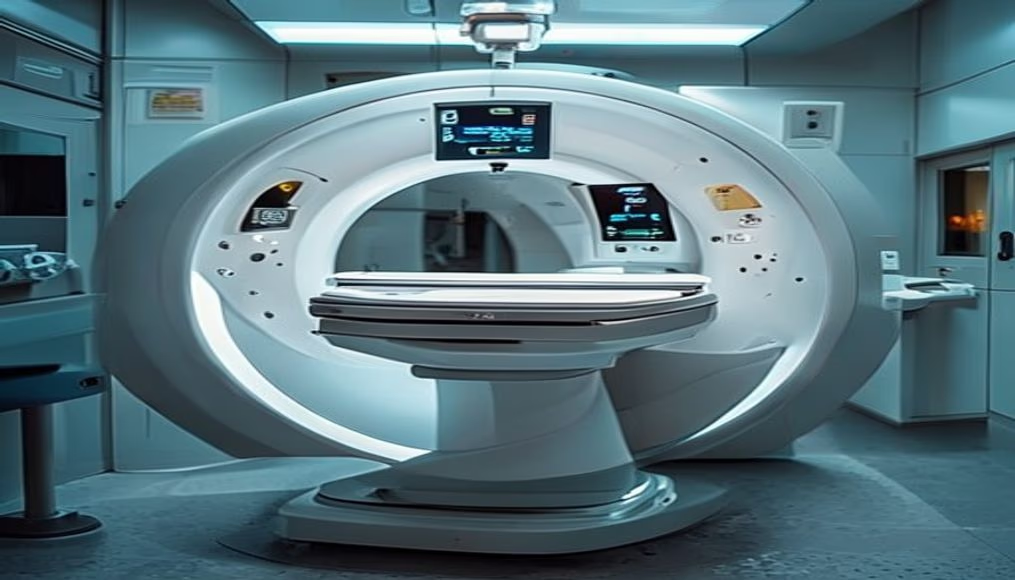
Mri scanner machine in radiology department
1344×768 · AVIF · CC BY 4.0
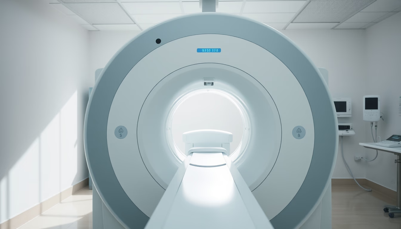
An MRI scanner machine in operation at a radiology department, essential equipment for non-invasive diagnostic imaging.
About this subject
Magnetic resonance imaging (MRI) is a medical imaging technique that uses strong magnetic fields and radio waves to generate detailed images of internal organs and tissues. Unlike X-rays or CT scans, MRI does not involve ionizing radiation, making it a safe tool for repeated examinations, especially in children and pregnant women. The equipment, like the one shown in the image, is a superconducting magnet that creates a uniform magnetic field, aligning hydrogen protons in the body. By applying radiofrequency pulses, these protons emit signals that are captured and processed by computers to form high-resolution images.
Hospital radiology departments house these scanners in rooms shielded against electromagnetic interference. The machine, with its central tunnel, allows examination from the brain to joints, being crucial for diagnosing tumors, spinal cord injuries, heart diseases, and neurological disorders such as multiple sclerosis. In Brazil, the Unified Health System (SUS) has over 500 MRI machines distributed across public and affiliated hospitals, although demand still outstrips supply in remote regions.
Interestingly, MRI development began in the 1970s, with scientists Paul Lauterbur and Peter Mansfield winning the Nobel Prize in Medicine in 2003. Since then, technology has evolved to faster and open scanners, reducing discomfort for claustrophobic patients. The symmetric image illuminated by natural light suggests a modern and well-designed environment, reflecting the importance of infrastructure for diagnostic accuracy.
Maintenance of these machines is complex and expensive, requiring specialized technicians. Liquid helium, used to cool superconducting magnets, needs periodic replenishment. Despite costs, MRI is indispensable in contemporary medicine, offering functional 3D images that guide surgeries and cancer treatments with millimeter precision.
Frequently Asked Questions
What is the difference between MRI and CT scan?
MRI uses magnetic fields and radio waves without ionizing radiation, ideal for soft tissues. CT uses X-rays, better for bones and emergencies.
Is it safe to have an MRI during pregnancy?
Yes, MRI is considered safe during pregnancy, especially in the second and third trimesters, as it does not use radiation. However, gadolinium contrast is avoided.
How long does an MRI scan take?
The time ranges from 15 to 60 minutes, depending on the area being examined and the need for contrast. Brain or spine scans are faster than multiple joint exams.
Direct URL
https://pub-c7d6a6ea828543ac903a74a341ccb2e1.r2.dev/imagens/mri-scanner-machine-in-radiology-department-symmetric-composition-bright-midday-sun.avifHow to credit
Include a visible link back to UtilizAí. Copy one of the snippets below:
<a href="https://xn--utiliza-eza.com/en/midia/imagens/mri-scanner-machine-in-radiology-department-symmetric-composition-bright-midday-sun">Mri scanner machine in radiology department</a> by <a href="https://xn--utiliza-eza.com">UtilizAí</a>, licensed under <a href="https://creativecommons.org/licenses/by/4.0/">CC BY 4.0</a>.
[Mri scanner machine in radiology department](https://xn--utiliza-eza.com/en/midia/imagens/mri-scanner-machine-in-radiology-department-symmetric-composition-bright-midday-sun) by [UtilizAí](https://xn--utiliza-eza.com), CC BY 4.0
License: CC-BY-4.0