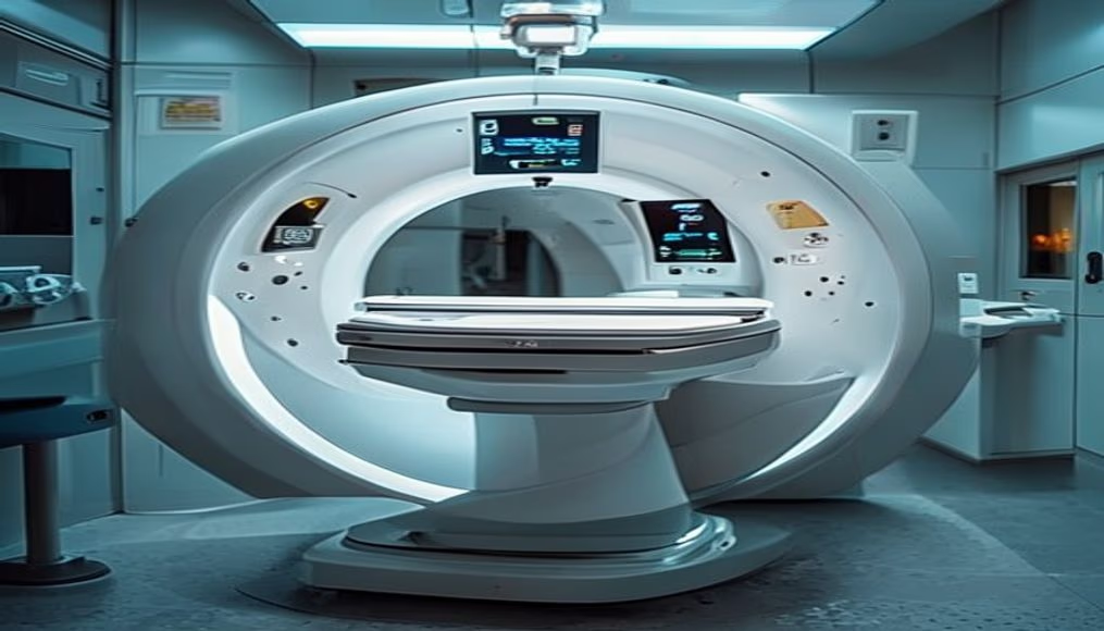
Mri scanner machine in radiology department
1344×768 · AVIF · CC BY 4.0
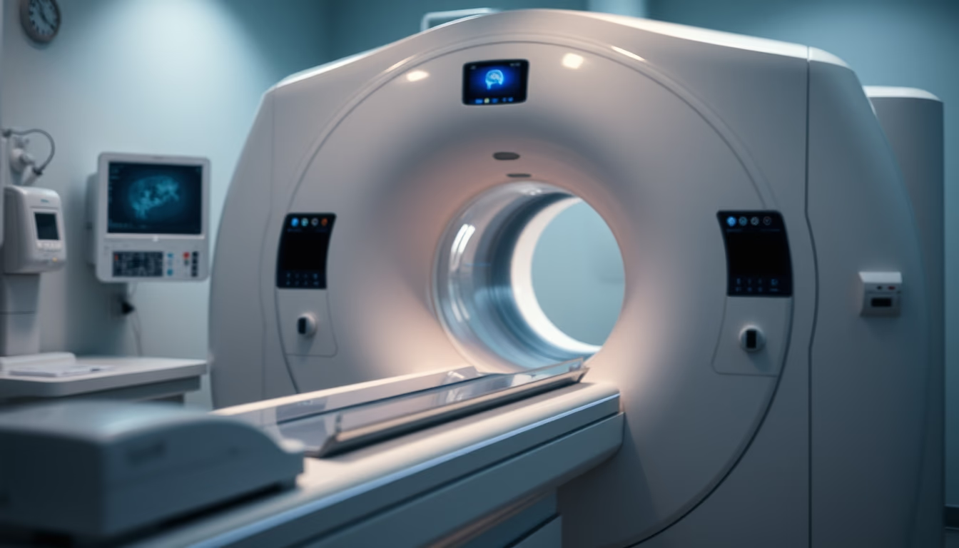
MRI scanner machine operating in a modern radiology department, essential for accurate diagnoses.
About this subject
Magnetic resonance imaging (MRI) is a non-invasive medical imaging technique that uses magnetic fields and radio waves to produce detailed images of organs and tissues. Unlike computed tomography, MRI does not use ionizing radiation, making it particularly safe for repeated examinations. The equipment, as shown, consists of a large tubular magnet where the patient is positioned. During the scan, the magnetic field aligns protons in hydrogen atoms within the body, and radiofrequency pulses are applied to disturb this alignment. As protons return to their original state, they emit signals that are captured and converted into high-resolution images.
The technology was developed starting in the 1970s, with the first human scan performed in 1977. Since then, it has become indispensable in neurology, orthopedics, and oncology, allowing visualization of tumors, spinal cord injuries, joint problems, and brain anomalies with clarity. The radiology department where the machine is installed follows strict safety protocols, including patient screening for metal objects or incompatible implants. The magnetic field is so intense that it can displace ferromagnetic objects, posing risks if not handled carefully.
In Brazil, MRI is available in public and private hospitals, but access remains unequal. The public health system (SUS) offers the exam, but waiting lists can be long in underserved regions. The machine in the image represents a high-field equipment (1.5 to 3 Tesla), common in reference centers. Maintenance and calibration are critical for image quality, involving specialized technicians. Recent advances include faster sequences and functional MRI, which maps brain activity in real time, useful in neurosurgery and cognitive research.
Frequently Asked Questions
Does an MRI hurt?
No, the exam is painless. The patient must lie still inside the machine, which may cause discomfort or claustrophobia in some individuals.
How long does an MRI scan take?
Typically 30 to 60 minutes, depending on the area being scanned and whether contrast is used.
Who cannot have an MRI?
People with pacemakers, non-compatible metal implants, aneurysm clips, or metal fragments in the body should avoid MRI. Screening is mandatory.
Direct URL
https://pub-c7d6a6ea828543ac903a74a341ccb2e1.r2.dev/imagens/mri-scanner-machine-in-radiology-department-documentary-photography-studio-professional-lighting.avifHow to credit
Include a visible link back to UtilizAí. Copy one of the snippets below:
<a href="https://xn--utiliza-eza.com/en/midia/imagens/mri-scanner-machine-in-radiology-department-documentary-photography-studio-professional-lighting">Mri scanner machine in radiology department</a> by <a href="https://xn--utiliza-eza.com">UtilizAí</a>, licensed under <a href="https://creativecommons.org/licenses/by/4.0/">CC BY 4.0</a>.
[Mri scanner machine in radiology department](https://xn--utiliza-eza.com/en/midia/imagens/mri-scanner-machine-in-radiology-department-documentary-photography-studio-professional-lighting) by [UtilizAí](https://xn--utiliza-eza.com), CC BY 4.0
License: CC-BY-4.0