Mri scanner machine in radiology department
1344×768 · AVIF · CC BY 4.0
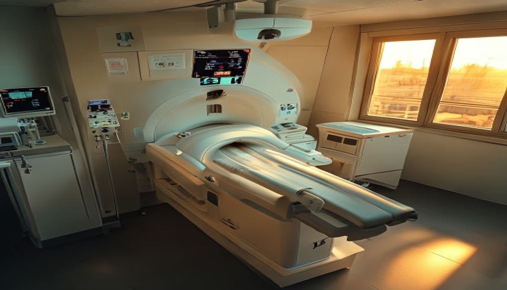
MRI scanner machine operating in a hospital radiology department, captured in an aerial shot during the late afternoon golden hour.
About this subject
Magnetic resonance imaging (MRI) is a non-invasive medical imaging technique that uses magnetic fields and radio waves to generate detailed images of internal organs and tissues. Unlike X-rays or CT scans, MRI does not use ionizing radiation, making it safer for repeated exams, especially in children and pregnant women. The equipment, such as the one shown in the image, consists of a large cylindrical magnet that creates an intense magnetic field, typically between 1.5 and 3 teslas. During the exam, the patient lies on a table that slides into the scanner, and the machine emits radiofrequency pulses that excite hydrogen atoms in the body, generating signals captured by a computer to form images.
In Brazil, MRI is widely used in public and private hospitals to diagnose conditions such as brain tumors, spinal cord injuries, joint disorders, and heart problems. According to the Ministry of Health, the country has over 3,000 MRI machines installed, mostly in metropolitan areas. The radiology department, where the machine is located, is a controlled environment with special shielding to prevent external interference with the magnetic field. The aerial shot captured during the golden hour, with late afternoon golden light, highlights the imposing equipment and the organization of the hospital space.
Interestingly, MRI was developed from nuclear magnetic resonance (NMR) studies in the 1940s, which earned the Nobel Prize in Physics for Felix Bloch and Edward Purcell. The first human body MRI image was obtained in 1977 by physician Raymond Damadian. Since then, technology has evolved rapidly, with faster scanners and higher magnetic fields, such as 7 tesla machines used in research. Safety is crucial: metallic objects or electronic devices can become dangerous projectiles near the magnet, so every patient undergoes rigorous screening before the exam.
Frequently Asked Questions
What is the difference between MRI and CT scan?
MRI uses magnetic fields and radio waves without ionizing radiation, ideal for soft tissues like the brain and muscles. CT scan uses X-rays and is faster, suitable for bones and emergencies.
Is it safe to have an MRI with metal implants?
It depends on the implant type. Devices such as pacemakers, aneurysm clips, or metal prosthetics may be contraindicated due to the magnetic field. It is essential to inform the doctor and technician about any metal in the body before the exam.
How long does an MRI scan take?
The exam can last from 15 to 60 minutes, depending on the body area and complexity. The patient must remain still during image acquisition to avoid blurring.
Direct URL
https://pub-c7d6a6ea828543ac903a74a341ccb2e1.r2.dev/imagens/mri-scanner-machine-in-radiology-department-aerial-drone-shot-golden-hour-late-afternoon.avifHow to credit
Include a visible link back to UtilizAí. Copy one of the snippets below:
<a href="https://xn--utiliza-eza.com/en/midia/imagens/mri-scanner-machine-in-radiology-department-aerial-drone-shot-golden-hour-late-afternoon">Mri scanner machine in radiology department</a> by <a href="https://xn--utiliza-eza.com">UtilizAí</a>, licensed under <a href="https://creativecommons.org/licenses/by/4.0/">CC BY 4.0</a>.
[Mri scanner machine in radiology department](https://xn--utiliza-eza.com/en/midia/imagens/mri-scanner-machine-in-radiology-department-aerial-drone-shot-golden-hour-late-afternoon) by [UtilizAí](https://xn--utiliza-eza.com), CC BY 4.0
License: CC-BY-4.0