Electron microscope sample
1344×768 · AVIF · CC BY 4.0
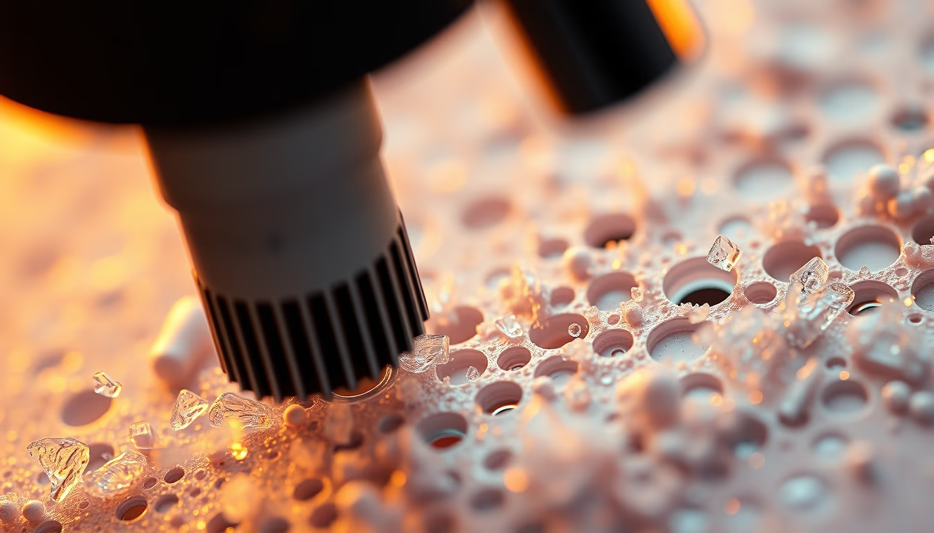
Electron microscope samples reveal ultrastructural details of cells and organisms, essential for advances in cell and molecular biology.
About this subject
Electron microscopy enables the observation of biological structures at nanometer scales, far beyond the reach of light microscopy. Samples undergo a rigorous preparation process: chemical or cryogenic fixation to preserve morphology, gradual dehydration, embedding in polymer resins, and sectioning into ultrathin slices (50-100 nanometers) using an ultramicrotome. In some cases, techniques such as freeze-fracture or cryo-substitution are employed to avoid ice crystal artifacts.
In biology, these samples are essential for studying cellular organelles (mitochondria, endoplasmic reticulum, Golgi apparatus), viruses, bacteria, and even macromolecules like proteins and DNA. Staining with heavy metals (uranium, lead, osmium) enhances contrast by scattering electrons, revealing details otherwise invisible. Routine pathological examinations use this method to diagnose kidney diseases, viral infections, and lysosomal storage disorders.
A classic example is the visualization of ribosomes attached to the rough endoplasmic reticulum, a discovery that revolutionized the understanding of protein synthesis. Today, cryo-electron microscopy (cryo-EM) won the Nobel Prize in Chemistry in 2017 for allowing determination of protein structures in their native state without crystallization. Careful sample preparation remains the most critical step for obtaining sharp and informative images.
Frequently Asked Questions
What is the typical thickness of a section for transmission electron microscopy?
Ultrathin sections are between 50 and 100 nanometers thick, about 1000 times thinner than a human hair.
Why do samples need to be stained with heavy metals?
Heavy metals (such as uranium and lead) increase contrast by strongly scattering electrons, making it possible to distinguish biological structures that have low natural electron density.
What is cryo-electron microscopy and how does it differ from conventional techniques?
Cryo-electron microscopy (cryo-EM) vitrifies the sample at cryogenic temperatures, preserving its natural hydration and avoiding artifacts from chemical fixation, allowing observation of molecules in near-native state.
Direct URL
https://pub-c7d6a6ea828543ac903a74a341ccb2e1.r2.dev/imagens/electron-microscope-sample-cinematic-wide-shot-p2.avifHow to credit
Include a visible link back to UtilizAí. Copy one of the snippets below:
<a href="https://xn--utiliza-eza.com/en/midia/imagens/electron-microscope-sample-cinematic-wide-shot-p2">Electron microscope sample</a> by <a href="https://xn--utiliza-eza.com">UtilizAí</a>, licensed under <a href="https://creativecommons.org/licenses/by/4.0/">CC BY 4.0</a>.
[Electron microscope sample](https://xn--utiliza-eza.com/en/midia/imagens/electron-microscope-sample-cinematic-wide-shot-p2) by [UtilizAí](https://xn--utiliza-eza.com), CC BY 4.0
License: CC-BY-4.0





