Cells under microscope
1344×768 · AVIF · CC BY 4.0
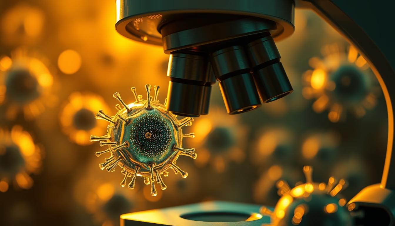
Human cells seen under a microscope reveal complex structures essential for life, such as the nucleus and organelles.
About this subject
Cells are the basic unit of life, and their observation under a microscope has revolutionized biology. Since Robert Hooke's first observations in 1665, which described cells in cork, to modern electron microscopes, the ability to visualize cellular structures has enabled advances in understanding diseases, drug development, and gene therapies. Typical human cells, such as fibroblasts, have a diameter between 10 and 100 micrometers and house organelles like mitochondria, endoplasmic reticulum, and the nucleus, which contains DNA. Staining techniques, such as hematoxylin and eosin, highlight different components, while fluorescence microscopy allows labeling of specific proteins. Cell study is fundamental to fields like oncology, immunology, and developmental biology, and microscope images are essential teaching and research tools in laboratories worldwide.
Frequently Asked Questions
What are cell organelles and what is their function?
Organelles are structures within the cell with specialized functions. For example, mitochondria produce energy, the endoplasmic reticulum synthesizes proteins and lipids, and the nucleus stores genetic material.
What is the difference between light and electron microscopy?
Light microscopy uses visible light to magnify up to about 1000x, while electron microscopy uses electron beams, achieving magnifications up to 1,000,000x, allowing visualization of organelles and even molecules.
How are cells stained for visualization?
Stains such as hematoxylin (a basic dye that marks nuclei) and eosin (an acidic dye that marks cytoplasm) are common. Immunofluorescence techniques use antibodies labeled with fluorophores for specific proteins.
Direct URL
https://pub-c7d6a6ea828543ac903a74a341ccb2e1.r2.dev/imagens/cells-under-microscope-cinematic-wide-shot-v2.avifHow to credit
Include a visible link back to UtilizAí. Copy one of the snippets below:
<a href="https://xn--utiliza-eza.com/en/midia/imagens/cells-under-microscope-cinematic-wide-shot-v2">Cells under microscope</a> by <a href="https://xn--utiliza-eza.com">UtilizAí</a>, licensed under <a href="https://creativecommons.org/licenses/by/4.0/">CC BY 4.0</a>.
[Cells under microscope](https://xn--utiliza-eza.com/en/midia/imagens/cells-under-microscope-cinematic-wide-shot-v2) by [UtilizAí](https://xn--utiliza-eza.com), CC BY 4.0
License: CC-BY-4.0