Antibody structure render
1344×768 · AVIF · CC BY 4.0
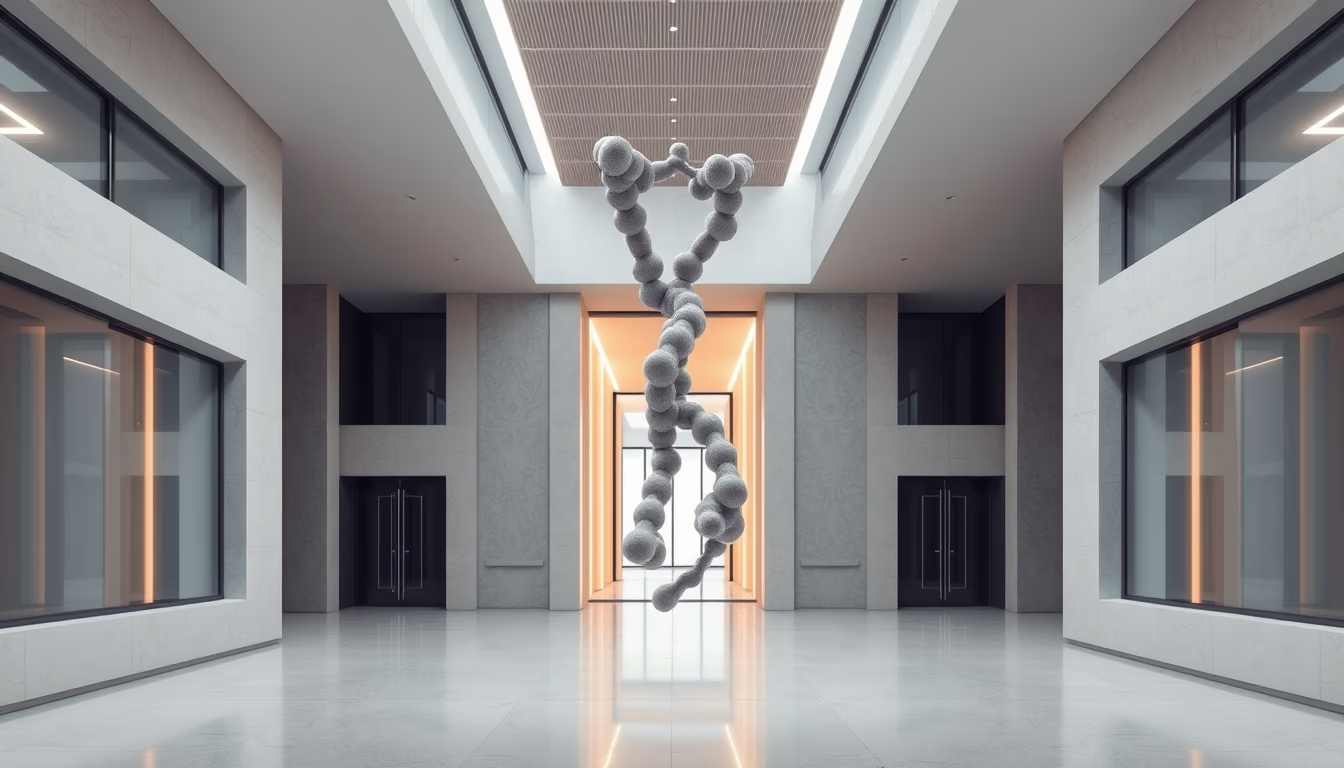
Renders of antibody structure reveal the molecular complexity of these proteins essential for the body's defense against pathogens.
About this subject
Antibodies, also known as immunoglobulins, are glycoproteins produced by B lymphocytes in response to the presence of antigens. Each antibody molecule has a Y-shaped structure composed of two heavy chains and two light chains linked by disulfide bonds. The variable regions at the tips of the Y are responsible for specific antigen recognition, while the constant region (Fc) determines the antibody class and interacts with other immune system cells.
There are five main classes of antibodies in mammals: IgM, IgG, IgA, IgD, and IgE. IgG is the most abundant in blood and tissues, crucial for acquired immunity. IgA predominates in mucous membranes and secretions such as breast milk, while IgE is involved in allergic responses and defense against parasites. The three-dimensional structure of these molecules has been elucidated by techniques such as X-ray crystallography, revealing details about antigen-binding sites.
Antibody engineering has revolutionized medicine, enabling the creation of therapeutic monoclonal antibodies for treating cancer, autoimmune diseases, and viral infections. Production involves fusing B cells with myeloma cells to generate hybridomas that secrete large quantities of identical antibodies. More recently, recombinant DNA techniques have allowed humanization of murine antibodies, reducing unwanted immune responses.
Visual representations of antibody structure, such as computational renders, aid researchers in understanding molecular interactions and designing new drugs. These images often highlight beta-sheets and alpha-helices of polypeptide chains, facilitating visualization of functional domains. In-depth study of antibody structure continues to drive discoveries in immunology and biotechnology.
Frequently Asked Questions
What is the main function of antibodies in the human body?
Antibodies recognize and bind to antigens from pathogens such as viruses and bacteria, neutralizing them or marking them for destruction by other immune cells.
How are monoclonal antibodies used in therapies produced?
Monoclonal antibodies are produced by hybridomas, hybrid cells formed by fusing a B lymphocyte with a myeloma cell, capable of secreting large quantities of a single type of antibody.
What distinguishes the different antibody classes (IgG, IgM, etc.)?
Classes differ in the constant region (Fc) structure and function: IgM is the first produced during infections, IgG is most abundant with long duration, IgA protects mucous membranes, IgE mediates allergies and parasite defense, and IgD acts as a receptor on B cells.
Direct URL
https://pub-c7d6a6ea828543ac903a74a341ccb2e1.r2.dev/imagens/antibody-structure-render-architectural-photography-p2.avifHow to credit
Include a visible link back to UtilizAí. Copy one of the snippets below:
<a href="https://xn--utiliza-eza.com/en/midia/imagens/antibody-structure-render-architectural-photography-p2">Antibody structure render</a> by <a href="https://xn--utiliza-eza.com">UtilizAí</a>, licensed under <a href="https://creativecommons.org/licenses/by/4.0/">CC BY 4.0</a>.
[Antibody structure render](https://xn--utiliza-eza.com/en/midia/imagens/antibody-structure-render-architectural-photography-p2) by [UtilizAí](https://xn--utiliza-eza.com), CC BY 4.0
License: CC-BY-4.0





