Protein structure 3d
1344×768 · AVIF · CC BY 4.0
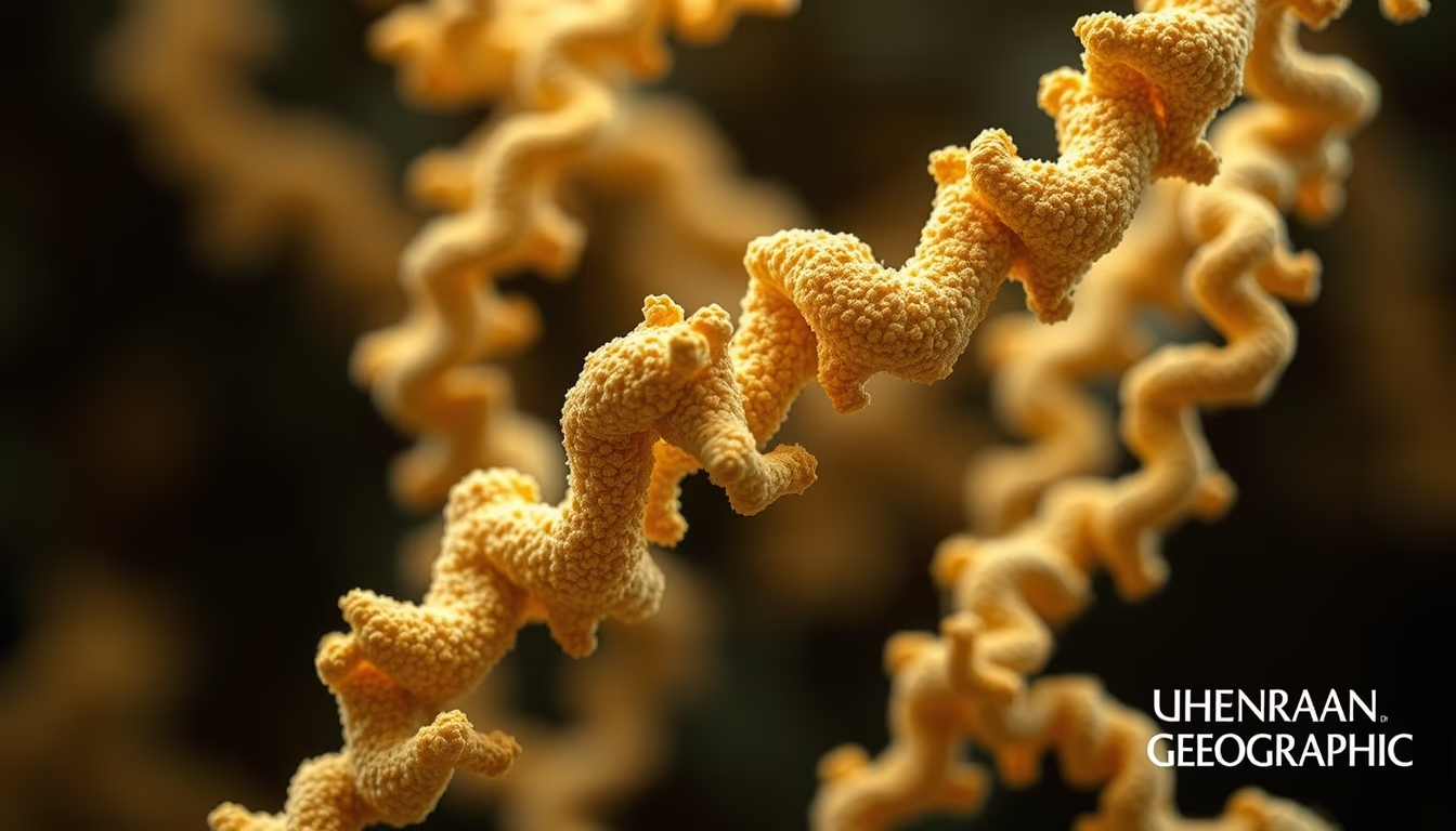
Discover how proteins organize into three-dimensional structures essential for life, from primary sequence to quaternary conformation.
About this subject
Proteins are complex molecules that perform vital functions in all living organisms, and their functionality depends directly on their three-dimensional structure. This structure is organized into four hierarchical levels: primary, secondary, tertiary, and quaternary. The primary level is the linear sequence of amino acids, determined by the genetic code. The secondary level involves local folding patterns such as α-helices and β-sheets, stabilized by hydrogen bonds. The tertiary level refers to the complete three-dimensional arrangement of a single polypeptide chain, including hydrophobic interactions, disulfide bridges, and ionic bonds. Finally, the quaternary level describes the assembly of multiple subunits, as in hemoglobin, which has four polypeptide chains.
Experimental determination of these structures revolutionized molecular biology. Techniques such as X-ray crystallography, nuclear magnetic resonance (NMR) spectroscopy, and cryo-electron microscopy (cryo-EM) allow visualization of atoms and interactions at atomic resolution. The Protein Data Bank (PDB) stores over 200,000 structures, accessible to researchers worldwide. Cryo-EM, in particular, has gained prominence by rendering dynamic and large molecular machines, such as ribosomes and ion channels.
The structure-function relationship is a central principle in biochemistry. A change in a single amino acid can alter folding and trigger diseases, like sickle cell anemia, where a substitution in hemoglobin leads to red blood cell deformation. Understanding the 3D structure of proteins also enables rational drug design, enzyme inhibitors, and therapeutic antibodies. For example, the development of HIV drugs was based on the structure of the viral protease.
In laboratories and classrooms, physical or digital three-dimensional models help students and scientists visualize abstract concepts. Tools like PyMOL and ChimeraX allow manipulation of structures, highlighting active sites and simulating interactions. Computational modeling, combined with artificial intelligence via AlphaFold, now predicts structures with accuracy comparable to experiments, accelerating discoveries in health and biotechnology.
Frequently Asked Questions
What determines the three-dimensional structure of a protein?
The three-dimensional structure is primarily determined by the amino acid sequence (primary level), which dictates how the polypeptide chain folds spontaneously, guided by chemical interactions such as hydrogen bonds, hydrophobic effects, and disulfide bridges. Environmental conditions like pH and temperature also influence folding.
How does protein structure affect its function?
The three-dimensional shape creates specific active sites and binding regions. For example, enzymes have cavities that fit substrates; antibodies recognize antigens. A structural change can inhibit function or cause malfunction, leading to diseases such as Alzheimer's, associated with misfolding of the beta-amyloid protein.
What is the most common technique for visualizing protein structures in 3D?
X-ray crystallography was historically the most common, but cryo-electron microscopy (cryo-EM) has become widely used for large complexes and membrane proteins. Nuclear magnetic resonance (NMR) is useful for small, dynamic proteins. Recently, computational prediction with AlphaFold has gained prominence.
Direct URL
https://pub-c7d6a6ea828543ac903a74a341ccb2e1.r2.dev/imagens/protein-structure-3d-national-geographic-style-p9.avifHow to credit
Include a visible link back to UtilizAí. Copy one of the snippets below:
<a href="https://xn--utiliza-eza.com/en/midia/imagens/protein-structure-3d-national-geographic-style-p9">Protein structure 3d</a> by <a href="https://xn--utiliza-eza.com">UtilizAí</a>, licensed under <a href="https://creativecommons.org/licenses/by/4.0/">CC BY 4.0</a>.
[Protein structure 3d](https://xn--utiliza-eza.com/en/midia/imagens/protein-structure-3d-national-geographic-style-p9) by [UtilizAí](https://xn--utiliza-eza.com), CC BY 4.0
License: CC-BY-4.0





