Protein structure 3d
1344×768 · AVIF · CC BY 4.0
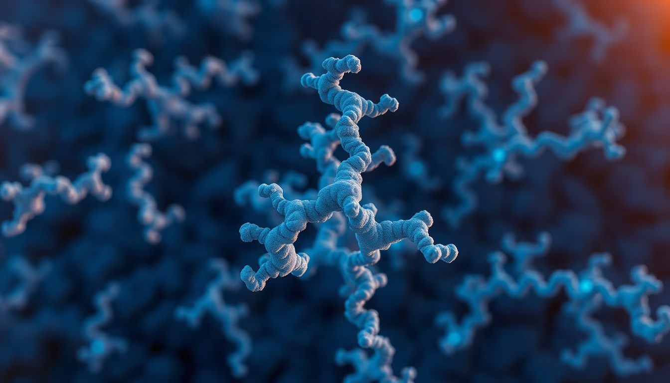
Three-dimensional protein structures reveal molecular conformations critical for biological functions such as catalysis and signaling.
About this subject
Proteins are macromolecules composed of amino acid chains that fold into complex three-dimensional structures. This conformation, known as tertiary structure, is determined by the amino acid sequence and interactions such as hydrogen bonds, hydrophobic interactions, and disulfide bridges. The 3D structure of a protein is fundamental to its function, defining active sites for catalysis, binding regions for other molecules, and regulatory mechanisms.
Methods such as X-ray crystallography, nuclear magnetic resonance (NMR) spectroscopy, and cryo-electron microscopy (cryo-EM) allow determination of protein atomic structures. X-ray crystallography, for example, diffracts X-rays from protein crystals to generate electron density maps, while cryo-EM analyzes frozen samples without crystallization. The Protein Data Bank (PDB) stores over 200,000 structures of proteins and other macromolecules, serving as a vital resource for structural biology research.
Understanding protein 3D structure has direct implications in medicine, such as drug development. An example is the modeling of inhibitors for viral proteins, such as HIV protease, which led to effective antiretroviral drugs. Additionally, protein engineering allows modification of structures to create industrial enzymes or therapeutics, such as monoclonal antibodies. Computational structure prediction, boosted by artificial intelligence like AlphaFold, has accelerated the field by predicting structures with high accuracy from the primary sequence.
Improper protein folding is associated with diseases such as Alzheimer's, Parkinson's, and mad cow disease, where protein aggregates (amyloids) accumulate. Studying these pathological structures aids in developing therapies that prevent misfolding or aggregation. In summary, analyzing protein 3D structure is a central tool in molecular biology, combining experiments and computational models to unravel the mechanisms of life.
Frequently Asked Questions
How is a protein's 3D structure determined experimentally?
Main methods include X-ray crystallography, nuclear magnetic resonance (NMR) spectroscopy, and cryo-electron microscopy (cryo-EM). X-ray crystallography requires protein crystals; NMR works on proteins in solution; cryo-EM analyzes frozen samples without crystallization.
What is the Protein Data Bank (PDB)?
The PDB is an international database that stores experimentally determined three-dimensional structures of proteins and nucleic acids. It is freely accessible and widely used by researchers for structural studies and drug design.
Why is AlphaFold important for protein structure prediction?
AlphaFold, developed by DeepMind, is an AI system that predicts a protein's 3D structure from its amino acid sequence with high accuracy, accelerating discoveries and reducing the need for expensive experimental methods.
Direct URL
https://pub-c7d6a6ea828543ac903a74a341ccb2e1.r2.dev/imagens/protein-structure-3d-aerial-drone-shot-p2.avifHow to credit
Include a visible link back to UtilizAí. Copy one of the snippets below:
<a href="https://xn--utiliza-eza.com/en/midia/imagens/protein-structure-3d-aerial-drone-shot-p2">Protein structure 3d</a> by <a href="https://xn--utiliza-eza.com">UtilizAí</a>, licensed under <a href="https://creativecommons.org/licenses/by/4.0/">CC BY 4.0</a>.
[Protein structure 3d](https://xn--utiliza-eza.com/en/midia/imagens/protein-structure-3d-aerial-drone-shot-p2) by [UtilizAí](https://xn--utiliza-eza.com), CC BY 4.0
License: CC-BY-4.0





