Nucleus chromosomes microscope
1344×768 · AVIF · CC BY 4.0
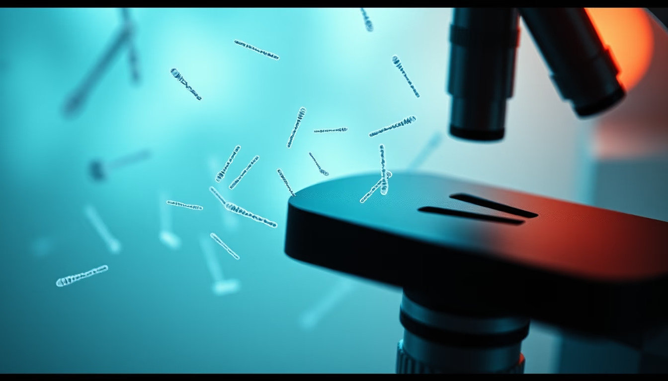
The cell nucleus houses chromosomes, structures that carry DNA and determine the hereditary traits of organisms.
About this subject
The nucleus is a membrane-bound organelle found in eukaryotic cells. It contains most of the cell's genetic material, organized into chromosomes. Each chromosome consists of a long DNA molecule associated with histone proteins, forming chromatin. During cell division, chromatin condenses to form chromosomes visible under a light microscope. Observing metaphase chromosomes enables cytogenetic studies, such as detecting numerical or structural abnormalities.
Light microscopy is a fundamental tool for visualizing chromosomes. Techniques like Giemsa staining produce characteristic banding patterns for each chromosome pair, allowing identification. The human karyotype, for example, consists of 23 chromosome pairs: 22 autosomes and one sex pair (XX or XY). Alterations in chromosome number or structure can be associated with syndromes like Down, Turner, and Klinefelter.
Beyond light microscopy, advanced techniques such as fluorescence in situ hybridization (FISH) and electron microscopy allow visualization of submicroscopic details. Chromosome study has implications in medicine, agriculture, and evolution. For instance, identifying chromosomal translocations is crucial in leukemia diagnosis. Modern genomic research uses chromosome DNA sequencing to map genes and understand hereditary diseases.
Frequently Asked Questions
What are chromosomes?
Chromosomes are structures within the cell nucleus that contain DNA, the molecule carrying genetic instructions. They become visible during cell division.
How many chromosomes do humans have?
Humans have 46 chromosomes, arranged in 23 pairs. One pair determines sex: XX for females, XY for males.
How are chromosomes studied?
Chromosomes are studied using techniques like karyotyping, which uses microscopy to analyze number and structure, and molecular methods such as FISH.
Direct URL
https://pub-c7d6a6ea828543ac903a74a341ccb2e1.r2.dev/imagens/nucleus-chromosomes-microscope-film-grain-authentic-p10.avifHow to credit
Include a visible link back to UtilizAí. Copy one of the snippets below:
<a href="https://xn--utiliza-eza.com/en/midia/imagens/nucleus-chromosomes-microscope-film-grain-authentic-p10">Nucleus chromosomes microscope</a> by <a href="https://xn--utiliza-eza.com">UtilizAí</a>, licensed under <a href="https://creativecommons.org/licenses/by/4.0/">CC BY 4.0</a>.
[Nucleus chromosomes microscope](https://xn--utiliza-eza.com/en/midia/imagens/nucleus-chromosomes-microscope-film-grain-authentic-p10) by [UtilizAí](https://xn--utiliza-eza.com), CC BY 4.0
License: CC-BY-4.0





