Nucleus chromosomes microscope
1344×768 · AVIF · CC BY 4.0
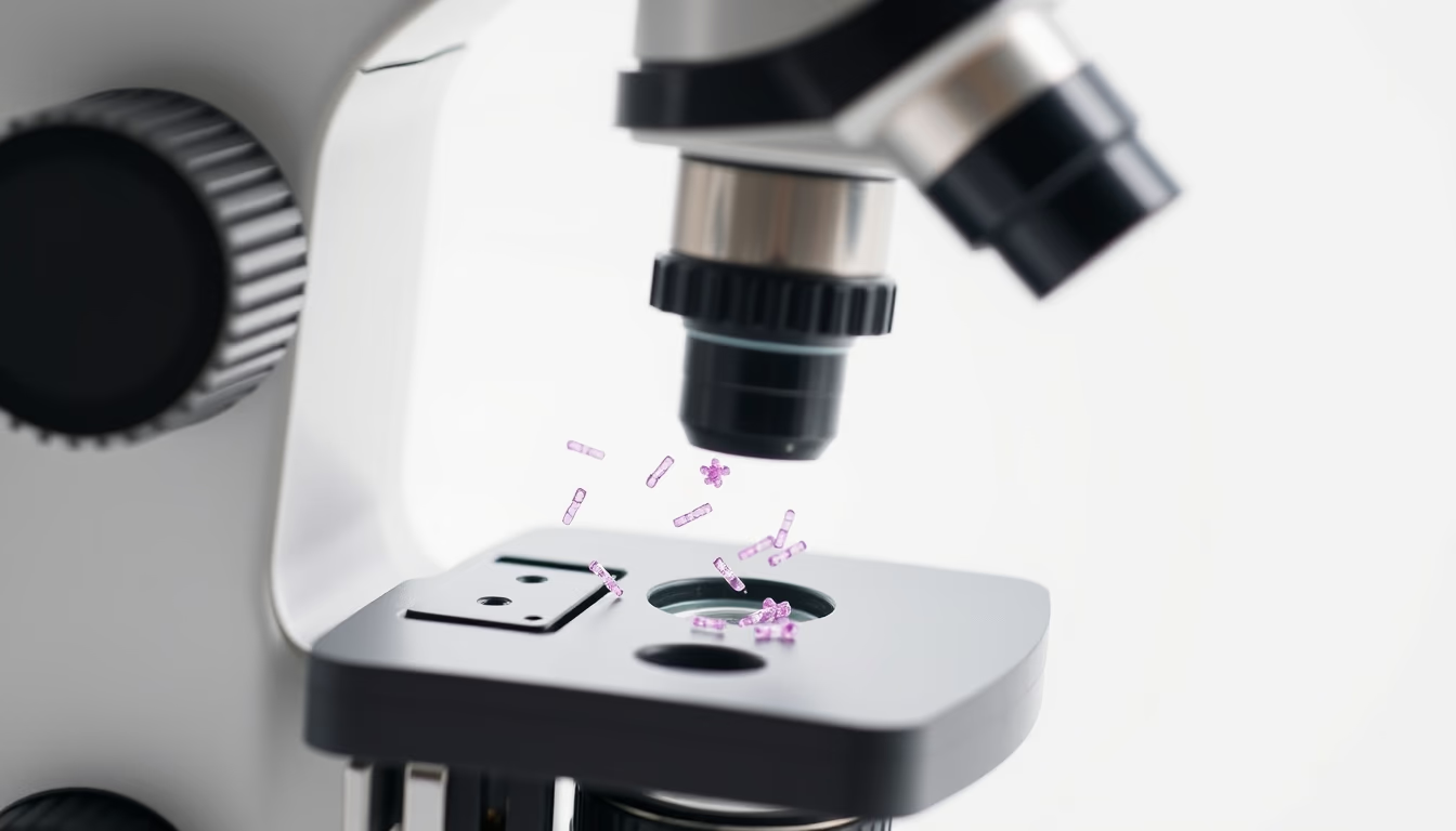
Chromosomes in the cell nucleus are structures that organize DNA and are visualized by microscopy for genetic studies.
About this subject
Chromosomes are thread-like structures made of DNA and histone proteins, located in the nucleus of eukaryotic cells. They carry the genetic information essential for organism development, function, and reproduction. During cell division, chromosomes condense and become visible under a light microscope, allowing scientists to study their morphology and number.
Microscopy of chromosomes is a fundamental tool in cytogenetics and genetic disease diagnosis. Techniques such as chromosome banding (e.g., G-banding, Q-banding) reveal unique band patterns, facilitating the identification of abnormalities like translocations, deletions, or duplications. These tests are often performed on peripheral blood cells, bone marrow, or fetal tissue.
In Brazil, cytogenetic services are provided by the public health system (SUS) for investigating syndromes such as Down, Turner, and Klinefelter. Chromosome analysis is also crucial in oncology, where chromosomal alterations are linked to various cancers. A typical human karyotype image shows 23 pairs of chromosomes: 22 autosomes and one sex pair (XY or XX).
Frequently Asked Questions
What are chromosomes made of?
Chromosomes are composed of a long DNA molecule associated with histone and non-histone proteins, forming chromatin. DNA carries the genes, while histones help compact the structure.
How are chromosomes visualized under a microscope?
For visualization, cells are cultured, treated with colchicine to arrest cell division at metaphase, and then stained with specific dyes. Chromosome banding reveals band patterns that allow identification of each chromosome.
What is the relationship between chromosomes and DNA?
Each chromosome contains a single linear DNA molecule that houses thousands of genes. DNA wraps around histones to form nucleosomes, which compact into chromosomes during cell division.
Direct URL
https://pub-c7d6a6ea828543ac903a74a341ccb2e1.r2.dev/imagens/nucleus-chromosomes-microscope-commercial-product-photography-p5.avifHow to credit
Include a visible link back to UtilizAí. Copy one of the snippets below:
<a href="https://xn--utiliza-eza.com/en/midia/imagens/nucleus-chromosomes-microscope-commercial-product-photography-p5">Nucleus chromosomes microscope</a> by <a href="https://xn--utiliza-eza.com">UtilizAí</a>, licensed under <a href="https://creativecommons.org/licenses/by/4.0/">CC BY 4.0</a>.
[Nucleus chromosomes microscope](https://xn--utiliza-eza.com/en/midia/imagens/nucleus-chromosomes-microscope-commercial-product-photography-p5) by [UtilizAí](https://xn--utiliza-eza.com), CC BY 4.0
License: CC-BY-4.0





