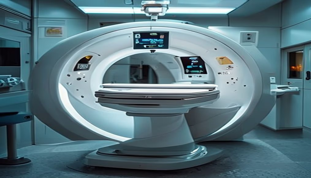
Mri scanner machine in radiology department
1344×768 · AVIF · CC BY 4.0
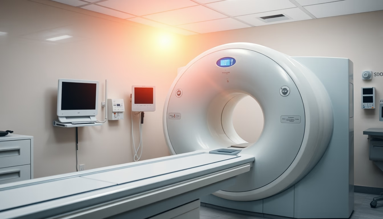
MRI scanner machine operating in a radiology department of a modern hospital.
About this subject
Magnetic resonance imaging (MRI) is a non-invasive medical imaging technique that uses magnetic fields and radio waves to generate detailed images of organs and tissues. Unlike computed tomography, MRI does not use ionizing radiation, making it safer for repeated scans, especially in children and pregnant women. The equipment consists of a large cylindrical magnet into which the patient is placed. During the exam, the magnetic field aligns hydrogen protons in the body, and radiofrequency pulses are applied to disrupt their alignment; as they return to their original state, they emit signals captured by antennas, which are processed by computer to form high-resolution three-dimensional images.
MRI is essential in diagnosing neurological, musculoskeletal, and oncological diseases. For example, it allows visualization of brain tumors, spinal cord injuries, ligament tears, and joint inflammation. It is also used to assess blood flow (MR angiography) and cardiac function. Technology has evolved to high-field systems (3 Tesla or more), offering greater clarity and reduced scan time. However, MRI has contraindications: patients with metallic implants (such as pacemakers, aneurysm clips, or ferromagnetic prostheses) cannot undergo the exam due to the risk of device displacement or heating.
Historically, MRI developed from nuclear magnetic resonance (NMR) studies by Bloch and Purcell in the 1940s, which earned the Nobel Prize in Physics in 1952. Medical application began in the 1970s with the work of Paul Lauterbur and Peter Mansfield, who also received the Nobel Prize in Medicine in 2003. Since then, MRI has become the gold standard for many clinical indications. In Brazil, the first machines were installed in the 1990s at university hospitals, and today the country has over 3,000 devices, mainly concentrated in the South and Southeast regions. The exam is covered by the Unified Health System (SUS) for priority cases, but demand still exceeds supply in many areas.
Frequently Asked Questions
How long does an MRI scan take?
Typically between 20 and 60 minutes, depending on the body part and protocol. Brain scans are faster, while full spine or joint exams may take longer.
Is any preparation required before an MRI?
Usually no fasting is needed, but you must remove metal objects (jewelry, watches, glasses) and inform the doctor about implants or claustrophobia. In some cases, fasting or intravenous contrast may be required.
Is MRI harmful to health?
No, because it does not use ionizing radiation. The magnetic field and radio waves are considered safe. However, patients with implanted metal devices or pregnant women in the first trimester should avoid the exam unless medically advised.
Direct URL
https://pub-c7d6a6ea828543ac903a74a341ccb2e1.r2.dev/imagens/mri-scanner-machine-in-radiology-department-documentary-photography-bright-midday-sun.avifHow to credit
Include a visible link back to UtilizAí. Copy one of the snippets below:
<a href="https://xn--utiliza-eza.com/en/midia/imagens/mri-scanner-machine-in-radiology-department-documentary-photography-bright-midday-sun">Mri scanner machine in radiology department</a> by <a href="https://xn--utiliza-eza.com">UtilizAí</a>, licensed under <a href="https://creativecommons.org/licenses/by/4.0/">CC BY 4.0</a>.
[Mri scanner machine in radiology department](https://xn--utiliza-eza.com/en/midia/imagens/mri-scanner-machine-in-radiology-department-documentary-photography-bright-midday-sun) by [UtilizAí](https://xn--utiliza-eza.com), CC BY 4.0
License: CC-BY-4.0