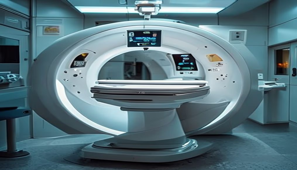
Mri scanner machine in radiology department
1344×768 · AVIF · CC BY 4.0
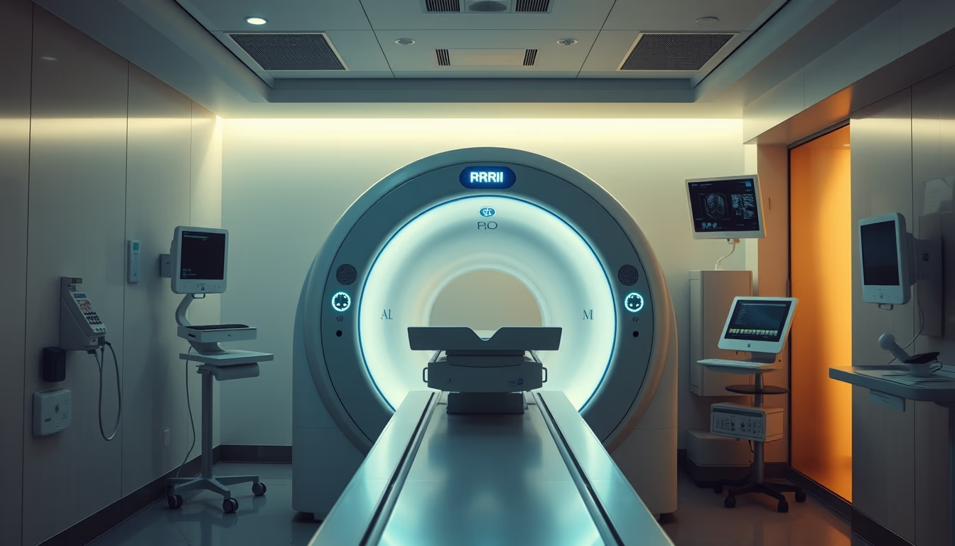
MRI scanner machine in a radiology department, an essential tool for non-invasive diagnostic imaging.
About this subject
Magnetic resonance imaging (MRI) is a medical imaging technique that uses strong magnetic fields and radio waves to generate detailed images of organs and tissues inside the body. Unlike X-rays or computed tomography (CT), MRI does not involve ionizing radiation, making it particularly safe for repeated exams and vulnerable populations such as children and pregnant women (though use during pregnancy is evaluated case by case). The equipment, as shown in the image, consists of a large tubular magnet where the patient lies on a table that slides into the scanner. During the exam, the machine emits radiofrequency pulses that excite hydrogen protons in the body's water molecules; as these protons return to their resting state, they release signals that are captured and processed by advanced computers to form images.
The history of MRI dates back to the 1970s, with pioneering work by Paul Lauterbur and Sir Peter Mansfield, who won the Nobel Prize in Medicine in 2003. The first clinical scan on a human was performed in 1977, and technology has since evolved rapidly. Modern scanners typically operate with magnetic field strengths of 1.5 to 3 Tesla (the unit of magnetic field measurement), while research-grade systems reach 7 Tesla or more, enabling increasingly higher resolution. In Brazil, MRI is available in both public and private hospitals, regulated by the National Health Surveillance Agency (ANVISA) for safety and quality standards.
The applications of MRI are vast: it is the preferred exam for evaluating the central nervous system (brain and spinal cord), joints (knee, shoulder, hip), heart and blood vessels, as well as the abdomen and pelvis. In oncology, MRI is essential for tumor staging and surgical planning. A curious fact: the loud, repetitive noise produced by the equipment during scanning is caused by the vibration of gradient coils when electrical current passes through them, which is why patients are given earplugs or headphones. Exam duration ranges from 20 to 60 minutes, depending on the region and complexity.
Safety is paramount: due to the extremely strong magnetic field, patients must remove all metal objects (jewelry, watches, credit cards), and those with implanted devices such as pacemakers, aneurysm clips, or metallic prostheses must be carefully screened. Radiology teams follow strict protocols to prevent accidents. The image of the scanner in a hospital setting with natural light reflects a modern, welcoming environment designed to reduce patient anxiety before the procedure.
Frequently Asked Questions
Does an MRI hurt or cause any discomfort?
The exam itself is painless, but it may cause discomfort due to the need to remain still for 20 to 60 minutes inside a confined space. Patients with claustrophobia may receive mild sedation. The loud noise from the machine is muffled with earplugs or headphones.
What are the contraindications for an MRI?
Absolute contraindications include patients with cardiac pacemakers, metallic cerebral aneurysm clips, cochlear implants, and intraocular metallic fragments. Devices such as stents, orthopedic prostheses, and IUDs are generally safe but must be evaluated by a physician.
What is the difference between MRI and CT scan?
MRI uses a magnetic field and radio waves, with no ionizing radiation, and is excellent for soft tissues (brain, muscles, cartilage). CT uses X-rays, is faster, and is better for evaluating bones, lungs, and emergencies like bleeding. The choice depends on the clinical indication.
Direct URL
https://pub-c7d6a6ea828543ac903a74a341ccb2e1.r2.dev/imagens/mri-scanner-machine-in-radiology-department-cinematic-wide-shot-morning-natural-light.avifHow to credit
Include a visible link back to UtilizAí. Copy one of the snippets below:
<a href="https://xn--utiliza-eza.com/en/midia/imagens/mri-scanner-machine-in-radiology-department-cinematic-wide-shot-morning-natural-light">Mri scanner machine in radiology department</a> by <a href="https://xn--utiliza-eza.com">UtilizAí</a>, licensed under <a href="https://creativecommons.org/licenses/by/4.0/">CC BY 4.0</a>.
[Mri scanner machine in radiology department](https://xn--utiliza-eza.com/en/midia/imagens/mri-scanner-machine-in-radiology-department-cinematic-wide-shot-morning-natural-light) by [UtilizAí](https://xn--utiliza-eza.com), CC BY 4.0
License: CC-BY-4.0