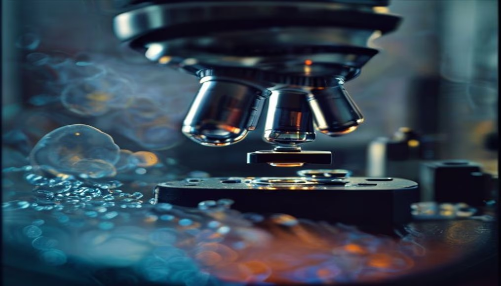
Lab technician looking through microscope
1344×768 · AVIF · CC BY 4.0
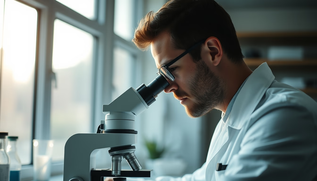
Lab technician analyzes samples under microscope during morning clinical research routine.
About this subject
Optical microscopy is a fundamental tool in health laboratories, enabling the visualization of cellular structures and microorganisms. The lab technician, a professional trained in clinical analysis, uses the microscope to examine slides with biological samples such as blood, urine, or tissues. This morning routine is common in university hospitals and research centers, where precision in identifying anomalies can impact diagnoses and treatments.
In Brazil, the Unified Health System (SUS) performs millions of microscopic exams per year, many in public laboratories. The training of these technicians includes subjects like histology, pathology, and biosafety. The use of binocular microscopes with magnification up to 1000x is standard, allowing the detection of parasites such as Plasmodium (the malaria causative agent) or cellular changes indicative of cancer.
Interestingly, modern microscopy began in the 17th century with Antonie van Leeuwenhoek, who first observed bacteria. Today, phase-contrast and fluorescence equipment expand possibilities, but the traditional optical microscope remains the first line of investigation in many Brazilian laboratories, especially in resource-limited regions.
Frequently Asked Questions
What training is required to become a lab technician in Brazil?
A lab technician needs a technical course in clinical analysis, lasting about two years, recognized by the Ministry of Education. Registration with the professional council (CRBM or CRF) is also required.
What types of samples are analyzed under a microscope?
The most common samples are blood, urine, feces, secretions, and tissue fragments. Each requires specific preparation, such as staining to highlight structures.
What is the difference between an optical and an electron microscope?
An optical microscope uses visible light and glass lenses, with a maximum magnification of about 1000x. An electron microscope uses electron beams, achieving magnifications up to 2 million times, ideal for viewing viruses and organelles.
Direct URL
https://pub-c7d6a6ea828543ac903a74a341ccb2e1.r2.dev/imagens/lab-technician-looking-through-microscope-documentary-morning-2.avifHow to credit
Include a visible link back to UtilizAí. Copy one of the snippets below:
<a href="https://xn--utiliza-eza.com/en/midia/imagens/lab-technician-looking-through-microscope-documentary-morning-2">Lab technician looking through microscope</a> by <a href="https://xn--utiliza-eza.com">UtilizAí</a>, licensed under <a href="https://creativecommons.org/licenses/by/4.0/">CC BY 4.0</a>.
[Lab technician looking through microscope](https://xn--utiliza-eza.com/en/midia/imagens/lab-technician-looking-through-microscope-documentary-morning-2) by [UtilizAí](https://xn--utiliza-eza.com), CC BY 4.0
License: CC-BY-4.0