Electron microscope sample
1344×768 · AVIF · CC BY 4.0
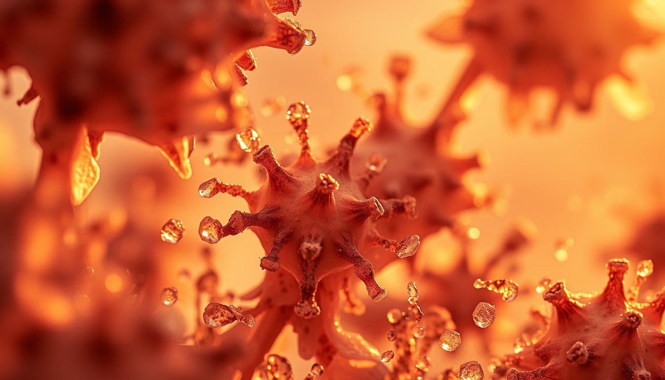
Samples for electron microscopy reveal details invisible to the naked eye, essential for studies in cell and molecular biology.
About this subject
Electron microscopy uses beams of electrons instead of light to achieve much higher resolution, allowing visualization of structures at the nanometer scale. Samples for this type of microscopy require specific preparation: dehydration, fixation, ultrathin sectioning (for TEM) or metal coating (for SEM). In biology, samples can include cells, tissues, viruses, and macromolecules. The technique was developed in the 1930s by Ernst Ruska, who won the Nobel Prize in Physics in 1986. Transmission electron microscopy (TEM) reveals the interior of cells, such as organelles and membranes, while scanning electron microscopy (SEM) generates three-dimensional images of sample surfaces. Proper preparation is crucial: poorly prepared samples can introduce artifacts or fail to withstand the vacuum chamber. Notably, recent advances allow observation of hydrated samples or under cryogenic conditions, as in cryo-electron microscopy, which earned the 2017 Nobel Prize in Chemistry for Jacques Dubochet, Joachim Frank, and Richard Henderson. This technique has revolutionized the determination of protein structures and molecular complexes.
Frequently Asked Questions
How are samples prepared for electron microscopy?
Samples undergo chemical fixation, dehydration, embedding in resin, ultrathin sectioning (for TEM), and coating with gold or carbon (for SEM) to conduct electrons and prevent charging.
What is the difference between transmission (TEM) and scanning (SEM) electron microscopy?
In TEM, the electron beam passes through the sample, revealing internal structures. In SEM, the beam scans the surface, producing three-dimensional topographic images.
Why is electron microscopy important in biology?
It allows visualization of organelles, viruses, and molecules like proteins and DNA at nanometer detail, essential for understanding cellular processes and diseases.
Direct URL
https://pub-c7d6a6ea828543ac903a74a341ccb2e1.r2.dev/imagens/electron-microscope-sample-travel-photography-p10.avifHow to credit
Include a visible link back to UtilizAí. Copy one of the snippets below:
<a href="https://xn--utiliza-eza.com/en/midia/imagens/electron-microscope-sample-travel-photography-p10">Electron microscope sample</a> by <a href="https://xn--utiliza-eza.com">UtilizAí</a>, licensed under <a href="https://creativecommons.org/licenses/by/4.0/">CC BY 4.0</a>.
[Electron microscope sample](https://xn--utiliza-eza.com/en/midia/imagens/electron-microscope-sample-travel-photography-p10) by [UtilizAí](https://xn--utiliza-eza.com), CC BY 4.0
License: CC-BY-4.0





