Electron microscope sample
1344×768 · AVIF · CC BY 4.0
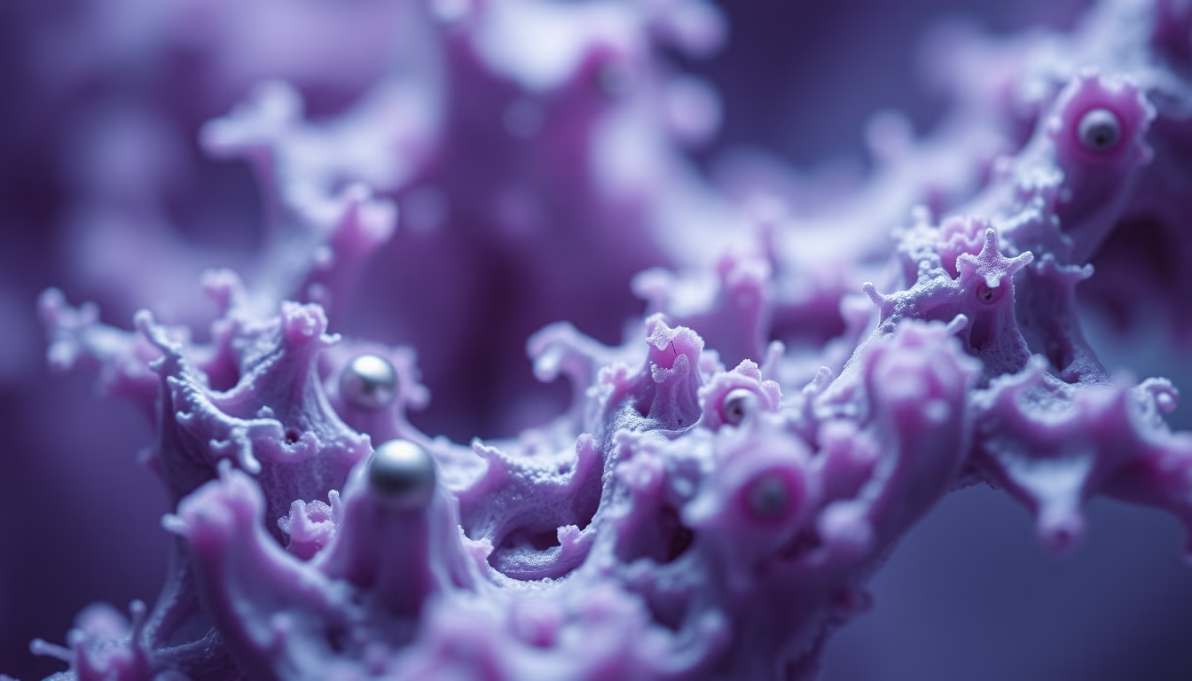
Electron microscopy enables visualization of structures at the nanometer scale, revealing details invisible to conventional light microscopes.
About this subject
Electron microscopy uses beams of electrons instead of light to illuminate samples, achieving magnifications of up to 10 million times. This resolution allows observation of cell organelles, viruses, and even individual atoms. The two main types are transmission electron microscopy (TEM) and scanning electron microscopy (SEM). In TEM, electrons pass through a thin sample, generating high-resolution internal images. SEM scans the surface with electrons, producing detailed three-dimensional images.
Sample preparation for electron microscopy is complex, requiring dehydration, chemical fixation, and often coating with gold or platinum for electrical conductivity. Biological samples must be cut into ultrathin slices 50 to 100 nanometers thick. This technique revolutionized cell biology, enabling the discovery of structures such as ribosomes, Golgi apparatus, and endoplasmic reticulum.
The development of the electron microscope began in the 1930s with Ernst Ruska and Max Knoll in Germany. Ruska received the Nobel Prize in Physics in 1986 for his invention. Modern electron microscopes can correct spherical and chromatic aberrations, achieving sub-angstrom resolution. Beyond biology, they are essential in materials science, semiconductors, and nanotechnology.
Trivia: The first commercial electron microscope was launched in 1939 by Siemens. Today, cutting-edge instruments cost millions of dollars and occupy climate-controlled rooms, requiring vacuum to prevent electron scattering by air molecules.
Frequently Asked Questions
What is the difference between transmission and scanning electron microscopes?
In TEM, electrons pass through the sample, revealing internal structure. In SEM, electrons scan the surface, generating three-dimensional images of topography.
How are biological samples prepared for electron microscopy?
Samples are chemically fixed, dehydrated, embedded in resin, and cut into ultrathin slices. Then they are coated with gold or platinum for electrical conductivity.
What is the maximum resolution of a modern electron microscope?
Aberration-corrected microscopes can resolve details smaller than 0.05 nanometers, allowing visualization of individual atoms.
Direct URL
https://pub-c7d6a6ea828543ac903a74a341ccb2e1.r2.dev/imagens/electron-microscope-sample-tilt-shift-miniature-p0.avifHow to credit
Include a visible link back to UtilizAí. Copy one of the snippets below:
<a href="https://xn--utiliza-eza.com/en/midia/imagens/electron-microscope-sample-tilt-shift-miniature-p0">Electron microscope sample</a> by <a href="https://xn--utiliza-eza.com">UtilizAí</a>, licensed under <a href="https://creativecommons.org/licenses/by/4.0/">CC BY 4.0</a>.
[Electron microscope sample](https://xn--utiliza-eza.com/en/midia/imagens/electron-microscope-sample-tilt-shift-miniature-p0) by [UtilizAí](https://xn--utiliza-eza.com), CC BY 4.0
License: CC-BY-4.0





