Electron microscope sample
1344×768 · AVIF · CC BY 4.0
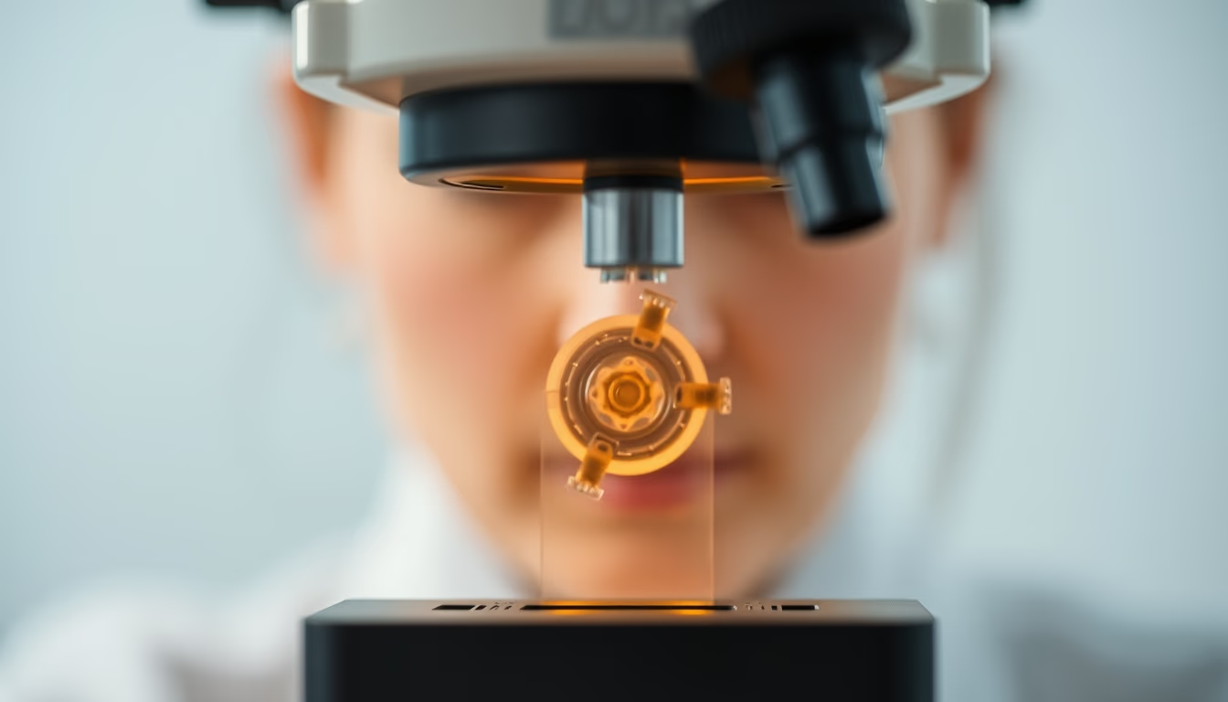
An electron microscope sample is prepared using specific techniques to reveal nanoscale structures, essential in biological and materials research.
About this subject
Samples for electron microscopy require meticulous preparation due to the high vacuum and electron beam. In biology, tissues are fixed, dehydrated, embedded in resin, and cut into ultrathin sections (50-100 nm) with an ultramicrotome. Heavy metals like uranium and lead are used for contrast. For materials, solid samples are polished or thinned by ion beam. Cryo-electron microscopy allows visualization of proteins in their native state by rapidly freezing the sample; this technique earned the 2017 Nobel Prize in Chemistry for Jacques Dubochet, Joachim Frank, and Richard Henderson. The choice of method depends on the goal: observing surfaces (SEM) or internal structure (TEM). In transmission electron microscopy, electrons pass through the sample, yielding atomic-resolution images. In scanning electron microscopy, a beam scans the surface, producing three-dimensional topography. Improper preparation can introduce artifacts like bubbles or ice crystals, compromising interpretation. Laboratories follow strict protocols to ensure reproducibility. In Brazil, institutions such as LNLS (National Synchrotron Light Laboratory) and USP provide advanced electron microscopy infrastructure, boosting research in virology, nanotechnology, and materials science.
Frequently Asked Questions
Why do biological samples need to be cut so thin for electron microscopy?
Electrons have low penetrating power, so samples must be less than 100 nanometers thick to allow transmission and achieve adequate resolution.
What is cryo-electron microscopy and how does it differ from conventional preparation?
In cryo-EM, the sample is rapidly frozen in liquid ethane, preserving its native structure without chemical fixation or stains, ideal for proteins and viruses.
What are the main artifacts in electron microscope sample preparation?
Common artifacts include air bubbles, ice crystals, shrinkage due to dehydration, and heavy metal deposits, which can distort the true morphology.
Direct URL
https://pub-c7d6a6ea828543ac903a74a341ccb2e1.r2.dev/imagens/electron-microscope-sample-studio-editorial-portrait-p3.avifHow to credit
Include a visible link back to UtilizAí. Copy one of the snippets below:
<a href="https://xn--utiliza-eza.com/en/midia/imagens/electron-microscope-sample-studio-editorial-portrait-p3">Electron microscope sample</a> by <a href="https://xn--utiliza-eza.com">UtilizAí</a>, licensed under <a href="https://creativecommons.org/licenses/by/4.0/">CC BY 4.0</a>.
[Electron microscope sample](https://xn--utiliza-eza.com/en/midia/imagens/electron-microscope-sample-studio-editorial-portrait-p3) by [UtilizAí](https://xn--utiliza-eza.com), CC BY 4.0
License: CC-BY-4.0





