Electron microscope sample
1344×768 · AVIF · CC BY 4.0
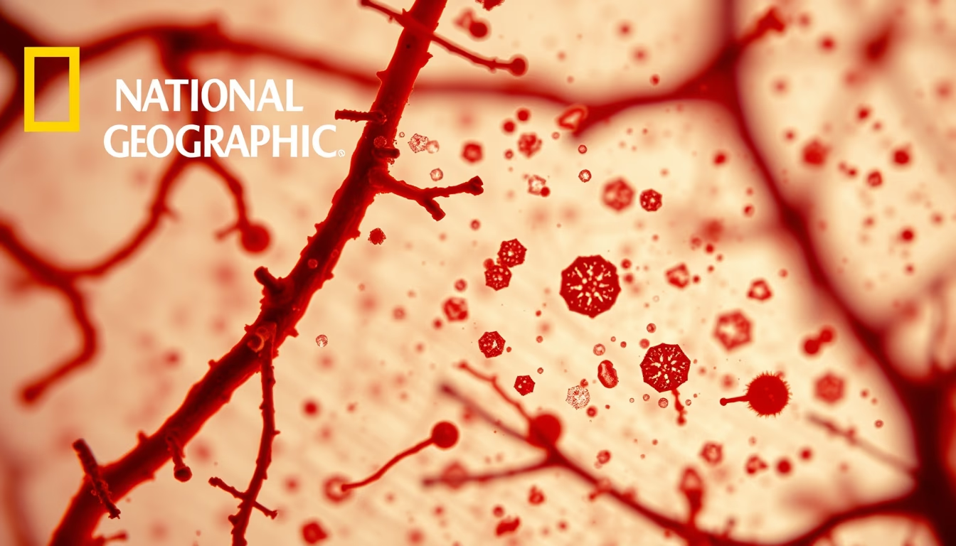
Electron microscopy unveils ultramicroscopic details of biological samples, revolutionizing cellular and microbial understanding.
About this subject
Electron microscopy represents a quantum leap in biological observation, surpassing the limitations of visible light. Unlike optical microscopes, which use photons, electron microscopes employ beams of electrons to illuminate the sample. This allows resolutions on the order of angstroms, enabling the visualization of viruses, cellular organelles, and macromolecules. The development of this instrument, initiated in the 1930s by Ernst Ruska and Max Knoll, earned Ruska the Nobel Prize in Physics in 1986. Since then, the technique has evolved into variants such as scanning electron microscopy (SEM) and transmission electron microscopy (TEM), each with specific applications.
Sample preparation for electron microscopy is a meticulous and critical process for obtaining clear images. Biological tissues must be chemically fixed with glutaraldehyde and osmium tetroxide, dehydrated through a series of alcohols, and embedded in hard resins like epoxy. Ultrathin sections, 50 to 100 nanometers thick, are cut with ultramicrotomes equipped with diamond knives. For SEM, samples must be dried at the critical point and coated with conductive metal, usually gold or platinum, to avoid electrical charge buildup.
Thanks to electron microscopy, fundamental discoveries have been made. The structure of DNA, the architecture of ribosomes, and the morphology of viruses such as HIV and SARS-CoV-2 were elucidated using these techniques. In botany, details of the cell wall and plasmodesmata were revealed. In microbiology, the presence of flagella and fimbriae in bacteria became evident. Today, cryo-electron microscopy allows observation of frozen samples in their native state without chemical fixation, opening new frontiers in structural biology. This technique was awarded the Nobel Prize in Chemistry in 2017.
Frequently Asked Questions
How does electron microscopy work?
It uses a beam of electrons focused by magnetic lenses to illuminate the sample. Electrons interact with the material, generating signals that are converted into an image. A vacuum is necessary to prevent electrons from being scattered by air molecules.
What types of samples can be observed?
Biological samples such as tissues, cells, viruses, and macromolecules, as well as materials like metals, ceramics, and polymers. They need to be conductive or coated with metal, and must be thin enough (for TEM) or vacuum-resistant (for SEM).
Why is electron microscopy important for biology?
It allows visualization of subcellular structures invisible to light microscopes, such as ribosomes, internal membranes, and protein complexes. This was essential for understanding processes like cell division, protein synthesis, and viral infection.
Direct URL
https://pub-c7d6a6ea828543ac903a74a341ccb2e1.r2.dev/imagens/electron-microscope-sample-national-geographic-style-p11.avifHow to credit
Include a visible link back to UtilizAí. Copy one of the snippets below:
<a href="https://xn--utiliza-eza.com/en/midia/imagens/electron-microscope-sample-national-geographic-style-p11">Electron microscope sample</a> by <a href="https://xn--utiliza-eza.com">UtilizAí</a>, licensed under <a href="https://creativecommons.org/licenses/by/4.0/">CC BY 4.0</a>.
[Electron microscope sample](https://xn--utiliza-eza.com/en/midia/imagens/electron-microscope-sample-national-geographic-style-p11) by [UtilizAí](https://xn--utiliza-eza.com), CC BY 4.0
License: CC-BY-4.0





