Electron microscope sample
1344×768 · AVIF · CC BY 4.0
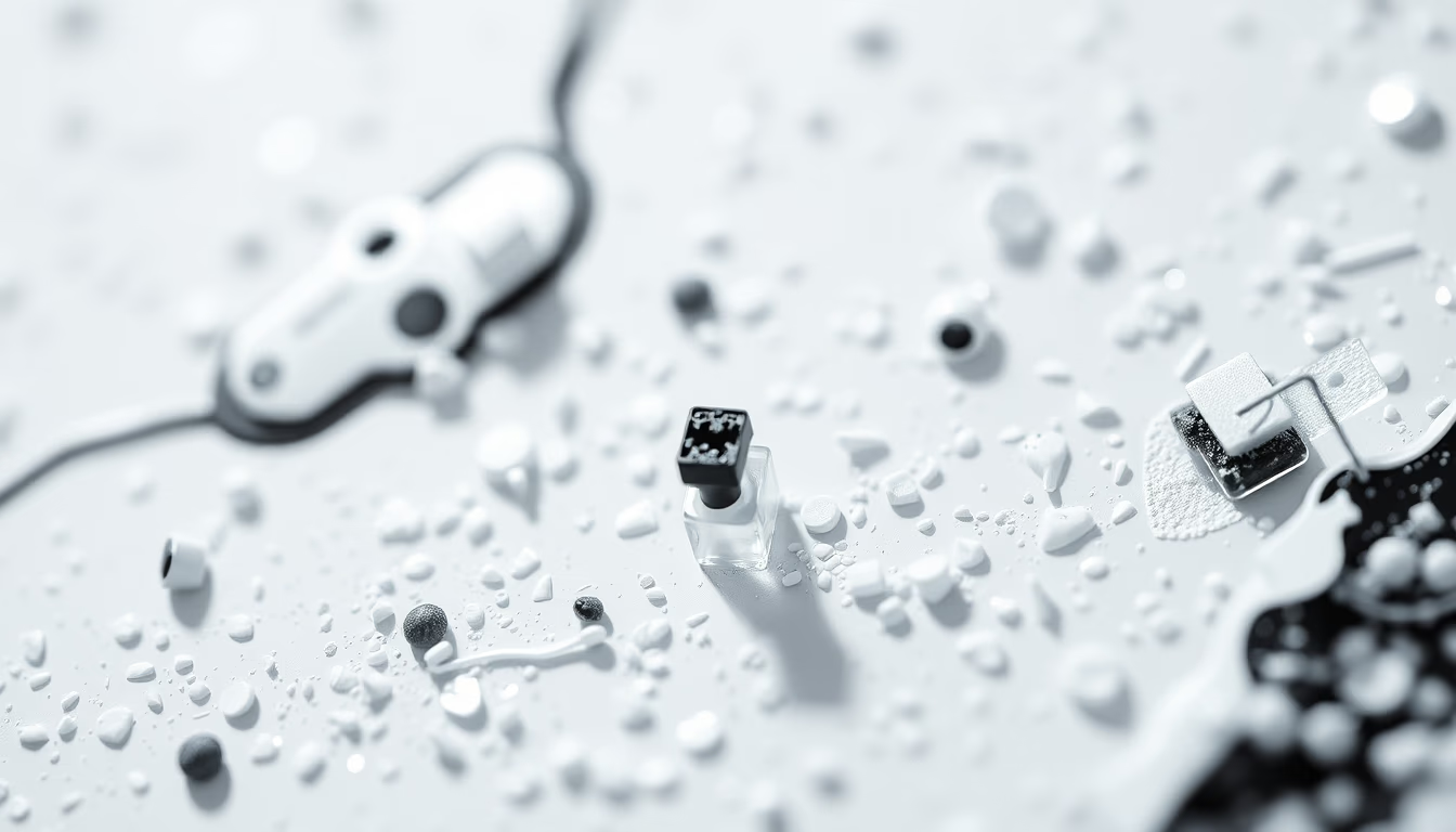
Samples for electron microscopes require meticulous preparation to reveal nanoscale structures, essential in cell and molecular biology.
About this subject
Electron microscopy revolutionized biology by enabling visualization of structures far beyond the reach of visible light. Biological samples such as cells, tissues, and viruses must undergo a rigorous preparation process to withstand the vacuum and electron beam. This process includes chemical fixation (using glutaraldehyde and osmium tetroxide), dehydration through graded ethanol or acetone series, and embedding in epoxy resins to produce ultrathin sections 50 to 100 nanometers thick.
For scanning electron microscopy (SEM), samples are critical-point dried and coated with a thin layer of gold or platinum to make them conductive. In transmission electron microscopy (TEM), ultrathin sections are stained with heavy metals like uranium and lead, which scatter electrons and generate detailed black-and-white images. These techniques reveal organelles such as mitochondria, endoplasmic reticulum, and Golgi complexes, as well as viral and protein structures.
The electron microscope was invented by Ernst Ruska in the 1930s, earning him the Nobel Prize in Physics in 1986. Since then, resolution has reached subnanometer scales, even allowing visualization of individual atoms in materials. In biology, cryo-electron microscopy (cryo-EM) has recently gained prominence because it avoids chemical fixation and allows observation of molecules in their native state by rapid freezing in liquid nitrogen.
Frequently Asked Questions
Why do samples need to be coated with gold or platinum in scanning electron microscopy?
The metal coating makes the sample conductive, preventing charge buildup that would distort the image and damage the material.
What is the difference between transmission electron microscopy (TEM) and scanning electron microscopy (SEM)?
In TEM, the electron beam passes through an ultrathin sample, forming images of internal structures. In SEM, the beam scans the surface, creating three-dimensional images of topography.
Has cryo-electron microscopy replaced traditional preparation methods?
Cryo-EM complements classical methods, especially for macromolecules and protein complexes, as it preserves native structure without fixation, but it still requires specialized equipment and frozen samples.
Direct URL
https://pub-c7d6a6ea828543ac903a74a341ccb2e1.r2.dev/imagens/electron-microscope-sample-minimalist-composition-p6.avifHow to credit
Include a visible link back to UtilizAí. Copy one of the snippets below:
<a href="https://xn--utiliza-eza.com/en/midia/imagens/electron-microscope-sample-minimalist-composition-p6">Electron microscope sample</a> by <a href="https://xn--utiliza-eza.com">UtilizAí</a>, licensed under <a href="https://creativecommons.org/licenses/by/4.0/">CC BY 4.0</a>.
[Electron microscope sample](https://xn--utiliza-eza.com/en/midia/imagens/electron-microscope-sample-minimalist-composition-p6) by [UtilizAí](https://xn--utiliza-eza.com), CC BY 4.0
License: CC-BY-4.0





