Cells under microscope
1344×768 · AVIF · CC BY 4.0
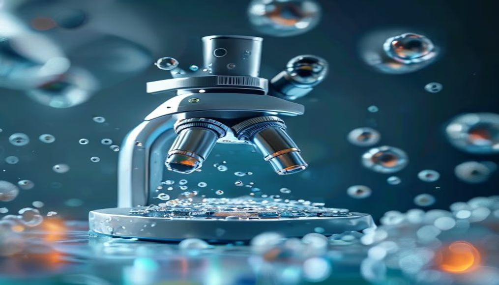
Cells observed under a microscope reveal the basic unit of life, essential for diagnostics and biomedical research.
About this subject
Cells are the smallest structural and functional unit of living organisms, capable of performing all life-sustaining activities. Since Robert Hooke's discovery in 1665, when he observed cork slices under a rudimentary microscope, the study of cells, cytology, has become a cornerstone of modern biology. There are prokaryotic cells, such as bacteria, without a defined nucleus, and eukaryotic cells, found in animals, plants, and fungi, with organelles like mitochondria and a membrane-bound nucleus.
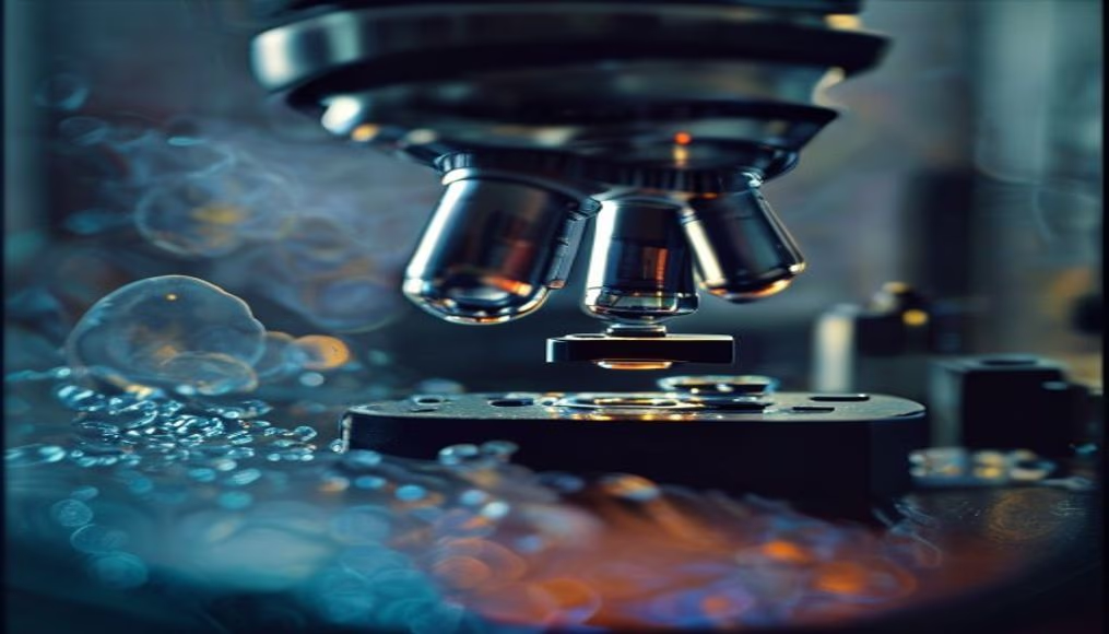
Microscopy has advanced immensely: from Antonie van Leeuwenhoek's early single-lens instruments, where he observed protozoa and spermatozoa, to modern scanning electron microscopes that can magnify up to 2 million times. Techniques like immunofluorescence and confocal microscopy allow visualization of specific proteins and intracellular structures in real time, revolutionizing understanding of processes such as cell division, signaling, and apoptosis.
In healthcare, cell analysis is critical for diagnosis. The Pap smear, for example, identifies abnormal cervical cells, preventing cancer. Complete blood counts measure red and white blood cells and platelets, indicating infections or anemias. Tissue biopsies, stained and examined under a microscope, help detect tumors and their characteristics. Additionally, cell culture is fundamental in developing vaccines and drugs, such as those against COVID-19.
Interestingly, an adult human body has about 37 trillion cells, with 2 billion dying and being replaced daily. Each cell contains the individual's complete DNA, but only a fraction of genes is activated depending on cell type. The study of stem cells, capable of differentiating into various tissues, opens prospects for regenerative therapies in diseases like Parkinson's and spinal cord injuries.
Frequently Asked Questions
What is a cell?
A cell is the smallest living unit of an organism, capable of growth, reproduction, and metabolic functions. All living beings are composed of one or more cells.
Who discovered cells and how?
English scientist Robert Hooke discovered cells in 1665 by examining thin slices of cork under a microscope. He observed small box-like structures and named them 'cells'.
Why is cell analysis under a microscope important in healthcare?
It helps diagnose diseases such as cancer, infections, and anemias, and monitors treatments. Tests like biopsies and complete blood counts rely on cell observation to detect pathological changes.
Direct URL
https://pub-c7d6a6ea828543ac903a74a341ccb2e1.r2.dev/imagens/cells-under-microscope-commercial-product-photography-v9.avifHow to credit
Include a visible link back to UtilizAí. Copy one of the snippets below:
<a href="https://xn--utiliza-eza.com/en/midia/imagens/cells-under-microscope-commercial-product-photography-v9">Cells under microscope</a> by <a href="https://xn--utiliza-eza.com">UtilizAí</a>, licensed under <a href="https://creativecommons.org/licenses/by/4.0/">CC BY 4.0</a>.
[Cells under microscope](https://xn--utiliza-eza.com/en/midia/imagens/cells-under-microscope-commercial-product-photography-v9) by [UtilizAí](https://xn--utiliza-eza.com), CC BY 4.0
License: CC-BY-4.0