Cell division microscope
1344×768 · AVIF · CC BY 4.0
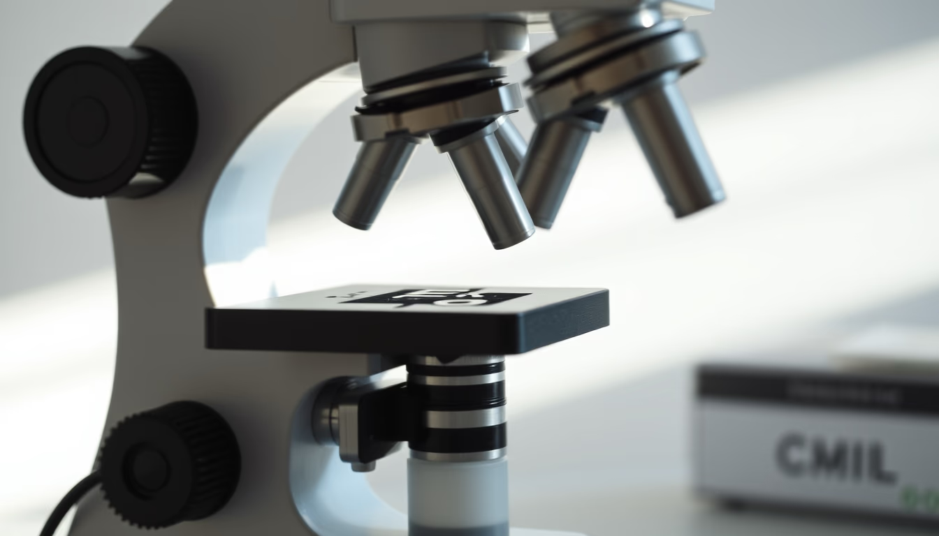
Cell division is the fundamental process by which cells reproduce, ensuring growth, repair, and continuity of life. Details observed under the microscope.
About this subject
Cell division is an essential process for life, allowing organisms to grow, repair damaged tissues, and reproduce. In eukaryotes, there are two main types: mitosis and meiosis. Mitosis produces two daughter cells genetically identical to the parent cell, crucial for development and cell renewal. Meiosis, on the other hand, generates four cells with half the genetic material, fundamental for sexual reproduction.
Under the microscope, the distinct phases of division can be observed: prophase, metaphase, anaphase, and telophase. In prophase, chromosomes condense and become visible as threadlike structures. In metaphase, they align at the cell's equator. Anaphase marks the separation of sister chromatids to opposite poles, and telophase sees the formation of two new nuclei. These stages were detailed by scientists such as Walther Flemming in the late 19th century.
Observing cell division under a microscope is a common practice in biology and medical labs. Stains like Giemsa or hematoxylin are often used to highlight chromosomes, allowing identification of abnormalities such as chromosomal changes associated with diseases. Studying mitosis and meiosis is also fundamental to understanding cancer, as failures in the regulation of cell division can lead to uncontrolled cell growth.
Frequently Asked Questions
What are the main differences between mitosis and meiosis?
Mitosis produces two daughter cells genetically identical to the parent, used for growth and repair. Meiosis yields four cells with half the genetic material, essential for gamete formation in sexual reproduction.
How does observing cell division help in disease diagnosis?
Chromosomal analysis during mitosis can reveal anomalies such as translocations or deletions, linked to genetic diseases and cancer. Karyotyping is a common diagnostic tool.
What is interphase in the cell cycle?
Interphase is the phase between two cell divisions, where the cell grows, replicates DNA, and prepares for mitosis or meiosis. It accounts for about 90% of the cell cycle.
Direct URL
https://pub-c7d6a6ea828543ac903a74a341ccb2e1.r2.dev/imagens/cell-division-microscope-minimalist-composition-p10.avifHow to credit
Include a visible link back to UtilizAí. Copy one of the snippets below:
<a href="https://xn--utiliza-eza.com/en/midia/imagens/cell-division-microscope-minimalist-composition-p10">Cell division microscope</a> by <a href="https://xn--utiliza-eza.com">UtilizAí</a>, licensed under <a href="https://creativecommons.org/licenses/by/4.0/">CC BY 4.0</a>.
[Cell division microscope](https://xn--utiliza-eza.com/en/midia/imagens/cell-division-microscope-minimalist-composition-p10) by [UtilizAí](https://xn--utiliza-eza.com), CC BY 4.0
License: CC-BY-4.0





