Cell division microscope
1344×768 · AVIF · CC BY 4.0
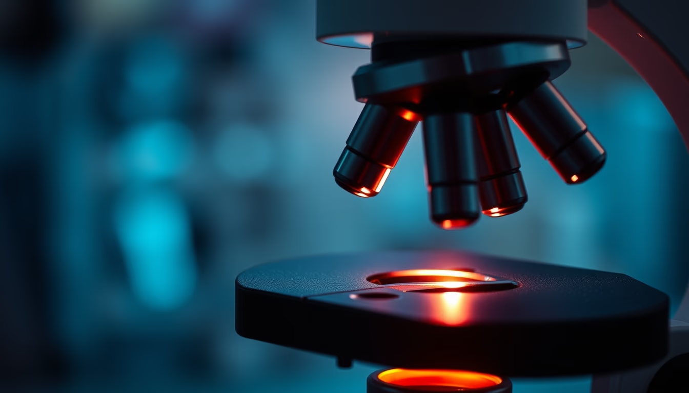
Cell division observed under the microscope reveals the fundamental mechanisms of growth and reproduction, from mitosis to meiosis.
About this subject
Cell division is the process by which a mother cell divides into two or more daughter cells, enabling growth, tissue repair, and reproduction. The two main types are mitosis, which produces genetically identical cells, and meiosis, which generates gametes with half the genetic material for sexual reproduction. Microscopic observation, pioneered by researchers like Walther Flemming in the 19th century, identified the phases of mitosis: prophase, metaphase, anaphase, and telophase.
Modern techniques such as fluorescence microscopy and live-cell time-lapse imaging allow real-time tracking of chromosome condensation, mitotic spindle formation, and cytokinesis. Electron microscopy reveals ultrastructural details like kinetochores and microtubules. These observations are vital for developmental biology, oncology, and cell therapy, as errors in cell division underlie diseases such as cancer.
The discovery of cell cycle control mechanisms, including cyclins and cyclin-dependent kinases, earned the 2001 Nobel Prize for Leland Hartwell, Tim Hunt, and Paul Nurse. Today, confocal and super-resolution microscopy enable visualization of subcellular structures at nanoscale, deepening our understanding of how cells reproduce precisely and orderly.
Frequently Asked Questions
What is the difference between mitosis and meiosis under the microscope?
In mitosis, a single nuclear division produces two daughter cells with the same chromosome number. In meiosis, two successive divisions yield four cells with half the chromosomes.
What microscopy techniques are used to study cell division?
Light microscopy with stains (e.g., DAPI for DNA), fluorescence microscopy with tagged proteins (GFP), live-cell time-lapse, confocal microscopy, and scanning/transmission electron microscopy.
Why is cell division important for medicine?
Understanding cell division helps explain tumor growth and develop chemotherapies that interrupt mitosis in cancer cells. It is also essential for regenerative therapies and assisted reproduction.
Direct URL
https://pub-c7d6a6ea828543ac903a74a341ccb2e1.r2.dev/imagens/cell-division-microscope-magazine-editorial-p12.avifHow to credit
Include a visible link back to UtilizAí. Copy one of the snippets below:
<a href="https://xn--utiliza-eza.com/en/midia/imagens/cell-division-microscope-magazine-editorial-p12">Cell division microscope</a> by <a href="https://xn--utiliza-eza.com">UtilizAí</a>, licensed under <a href="https://creativecommons.org/licenses/by/4.0/">CC BY 4.0</a>.
[Cell division microscope](https://xn--utiliza-eza.com/en/midia/imagens/cell-division-microscope-magazine-editorial-p12) by [UtilizAí](https://xn--utiliza-eza.com), CC BY 4.0
License: CC-BY-4.0





