Cell division microscope
1344×768 · AVIF · CC BY 4.0
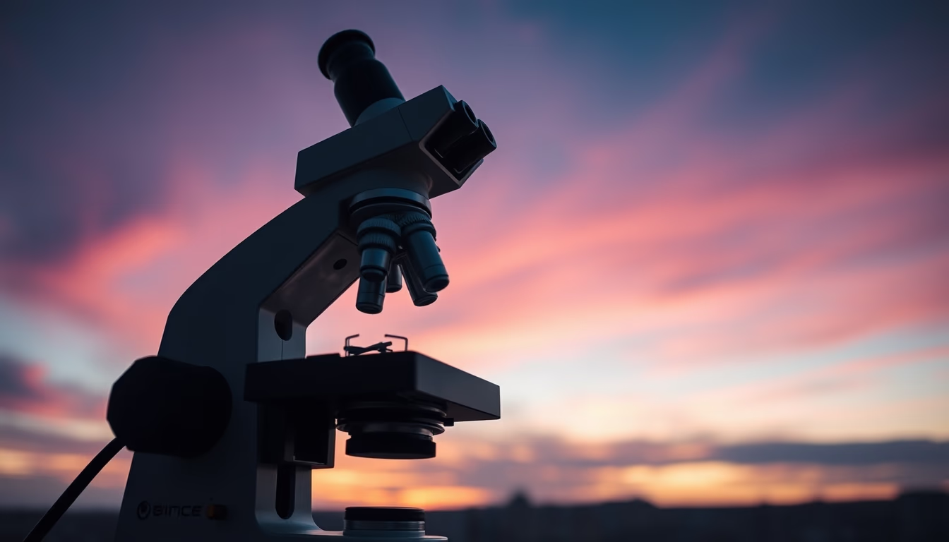
Cell division observed under a microscope: understand the process of mitosis and meiosis captured using long exposure technique.
About this subject
Cell division is a fundamental process for life, enabling growth, tissue repair, and reproduction. There are two main types: mitosis, which produces two identical daughter cells for growth and cell replacement, and meiosis, which generates gametes with half the genetic material, essential for sexual reproduction. Observing these processes under a microscope revolutionized biology, allowing 19th-century scientists like Walther Flemming to describe chromatin and karyokinesis.
To record the dynamics of cell division, imaging techniques such as long exposure are employed. Long exposure captures slow, continuous movements, like chromosome condensation and mitotic spindle formation, over minutes or hours. This requires stable optical systems and fluorescent markers such as GFP (green fluorescent protein), which illuminate specific structures without harming living cells.
Long-exposure microscopy reveals critical details, such as chromosome separation in anaphase and cytokinesis. These studies have practical applications in understanding diseases like cancer, where uncontrolled cell division is characteristic, and in developing therapies that halt mitosis in tumor cells. Today, advanced techniques like super-resolution fluorescence microscopy achieve nanometer scales, but traditional long exposure remains valuable for filming long cellular processes.
A notable example is the recording of HeLa cell division, an immortalized cell line used in research. Time-lapse videos combine long exposure with regular intervals, resulting in animations that show the full cell cycle. These resources are used in education and research, helping to visualize events in a living cell in an instructive and impactful way.
Frequently Asked Questions
What is mitosis and what are its phases?
Mitosis is the cell division process that produces two genetically identical daughter cells. Its phases are prophase, prometaphase, metaphase, anaphase, telophase, and cytokinesis.
How is long exposure used in cell microscopy?
Long exposure captures slow movements over minutes or hours, using stable illumination and fluorescent markers to record processes like chromosome condensation and segregation during division.
Why is cell division important in cancer research?
Cancer involves uncontrolled cell division; studying mitosis helps identify drug targets that block tumor cell proliferation, such as antimitotic agents.
Direct URL
https://pub-c7d6a6ea828543ac903a74a341ccb2e1.r2.dev/imagens/cell-division-microscope-long-exposure-p1.avifHow to credit
Include a visible link back to UtilizAí. Copy one of the snippets below:
<a href="https://xn--utiliza-eza.com/en/midia/imagens/cell-division-microscope-long-exposure-p1">Cell division microscope</a> by <a href="https://xn--utiliza-eza.com">UtilizAí</a>, licensed under <a href="https://creativecommons.org/licenses/by/4.0/">CC BY 4.0</a>.
[Cell division microscope](https://xn--utiliza-eza.com/en/midia/imagens/cell-division-microscope-long-exposure-p1) by [UtilizAí](https://xn--utiliza-eza.com), CC BY 4.0
License: CC-BY-4.0





