Cell division microscope
1344×768 · AVIF · CC BY 4.0
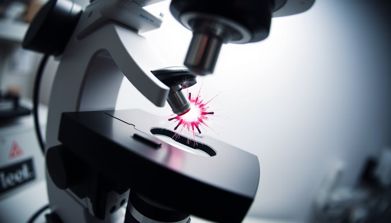
Cell division is a fundamental process for growth, repair, and reproduction of organisms, observed in detail through microscopy.
About this subject
Cell division is an essential biological process that allows tissue growth, cell renewal, and reproduction of organisms. In multicellular organisms such as humans, billions of cells divide daily to replace dead or damaged cells. There are two main types of cell division: mitosis and meiosis. Mitosis produces two daughter cells genetically identical to the mother cell and is responsible for growth and repair. Meiosis, which occurs in germ cells, generates four cells with half the genetic material, crucial for sexual reproduction.
Observing cell division under the microscope revolutionized biology. In the late 19th century, German biologist Walther Flemming used dyes to visualize chromosomes during mitosis, describing the stages of the process for the first time. Today, advanced techniques such as fluorescence microscopy and phase contrast allow real-time tracking of microtubule dynamics and chromosome separation. Specific dyes or stains like thymol can highlight structures such as the mitotic spindle.
The cell cycle is tightly controlled by proteins such as cyclins and cyclin-dependent kinases. Failures in this control can lead to tumor formation. In a healthy adult, approximately 5 million cells divide every second, a process invisible to the naked eye but revealed in all its complexity by microscopy. The fisheye lens effect in the image enhances the perception of this microscopic world, evoking the observer's immersion into the cellular universe.
Frequently Asked Questions
What is mitosis and why is it important?
Mitosis is the process of cell division that results in two identical daughter cells. It is essential for growth, tissue repair, and replacing dead cells.
How does microscopy allow observation of cell division?
Techniques such as staining, fluorescence microscopy, and phase contrast enable visualization of chromosomes, spindles, and other structures during cell division.
What is the difference between mitosis and meiosis?
Mitosis produces two genetically identical diploid cells; meiosis produces four genetically diverse haploid cells used in sexual reproduction.
Direct URL
https://pub-c7d6a6ea828543ac903a74a341ccb2e1.r2.dev/imagens/cell-division-microscope-fish-eye-wide-angle-p3.avifHow to credit
Include a visible link back to UtilizAí. Copy one of the snippets below:
<a href="https://xn--utiliza-eza.com/en/midia/imagens/cell-division-microscope-fish-eye-wide-angle-p3">Cell division microscope</a> by <a href="https://xn--utiliza-eza.com">UtilizAí</a>, licensed under <a href="https://creativecommons.org/licenses/by/4.0/">CC BY 4.0</a>.
[Cell division microscope](https://xn--utiliza-eza.com/en/midia/imagens/cell-division-microscope-fish-eye-wide-angle-p3) by [UtilizAí](https://xn--utiliza-eza.com), CC BY 4.0
License: CC-BY-4.0





