Ribosome detail render
1344×768 · AVIF · CC BY 4.0
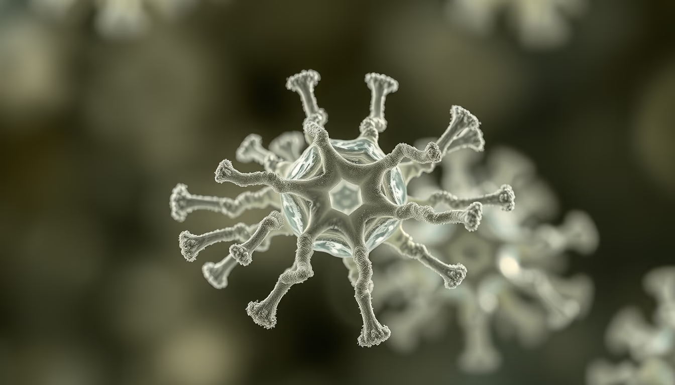
Ribosomes are molecular complexes responsible for protein synthesis in cells, translating messenger RNA into polypeptide chains.
About this subject
Ribosomes are fundamental molecular machines found in all living cells, from bacteria to humans. They are composed of two main subunits, large and small, which join during translation of messenger RNA (mRNA). The small subunit reads the genetic code of the mRNA, while the large subunit catalyzes the formation of peptide bonds between amino acids, building the protein. This process is known as protein synthesis and is essential for life.
Each ribosome is made of ribosomal RNA (rRNA) and proteins. In eukaryotic cells, ribosomes can be free in the cytoplasm or bound to the rough endoplasmic reticulum, synthesizing proteins that will be secreted or incorporated into membranes. In prokaryotes, ribosomes are smaller (70S) compared to eukaryotes (80S), a difference exploited by antibiotics such as tetracycline, which selectively inhibits bacterial ribosomes.
The discovery of ribosomes dates back to the early 1950s, with electron microscopy studies by George Palade, which earned him the Nobel Prize in Physiology or Medicine in 1974. Today, cryo-electron microscopy allows visualization of the atomic structure of ribosomes with stunning detail, revealing active sites and conformational movements. These advances drive the development of new antibiotics and the understanding of genetic diseases linked to translation defects.
Frequently Asked Questions
What is the main function of the ribosome?
The ribosome is responsible for protein synthesis, translating the genetic information in messenger RNA (mRNA) into a sequence of amino acids that forms a protein.
Why are ribosomes targets for antibiotics?
Antibiotics like tetracycline and erythromycin specifically target bacterial ribosomes (70S), inhibiting bacterial protein synthesis without affecting human ribosomes (80S), allowing treatment of infections.
How was the structure of the ribosome discovered?
The structure of the ribosome was first revealed by electron microscopy in the 1950s by George Palade. Today, techniques such as cryo-electron microscopy allow visualization of atomic-level details.
Direct URL
https://pub-c7d6a6ea828543ac903a74a341ccb2e1.r2.dev/imagens/ribosome-detail-render-magazine-editorial-p5.avifHow to credit
Include a visible link back to UtilizAí. Copy one of the snippets below:
<a href="https://xn--utiliza-eza.com/en/midia/imagens/ribosome-detail-render-magazine-editorial-p5">Ribosome detail render</a> by <a href="https://xn--utiliza-eza.com">UtilizAí</a>, licensed under <a href="https://creativecommons.org/licenses/by/4.0/">CC BY 4.0</a>.
[Ribosome detail render](https://xn--utiliza-eza.com/en/midia/imagens/ribosome-detail-render-magazine-editorial-p5) by [UtilizAí](https://xn--utiliza-eza.com), CC BY 4.0
License: CC-BY-4.0





