Protein structure 3d
1344×768 · AVIF · CC BY 4.0
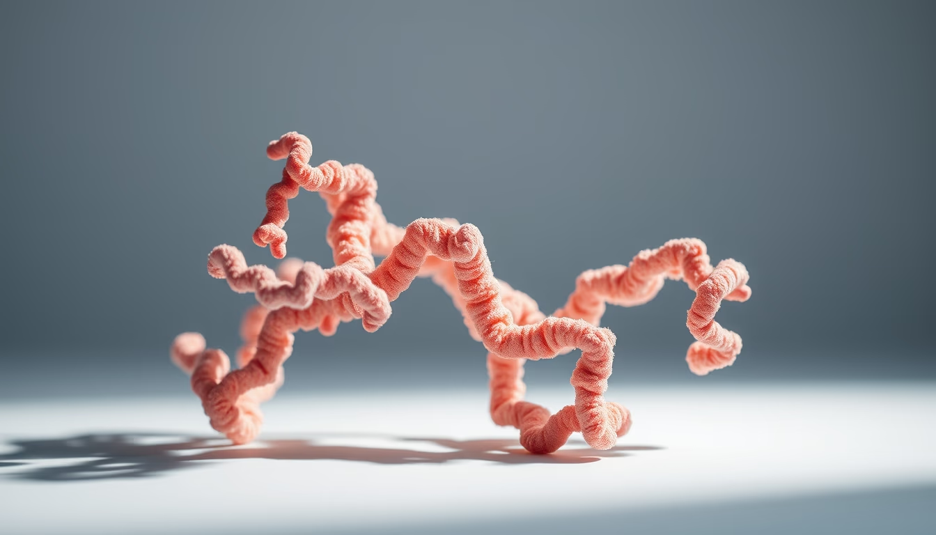
Proteins are essential macromolecules with complex three-dimensional structures that determine their biological functions.
About this subject
Proteins are composed of amino acid chains that fold into specific three-dimensional structures, classified into four levels: primary (linear sequence), secondary (alpha-helices and beta-sheets), tertiary (complete folding of a single chain), and quaternary (association of multiple chains). This 3D architecture is crucial for biological activity, as it determines binding sites and interactions with other molecules.
Experimental determination of protein structure employs techniques such as X-ray crystallography, nuclear magnetic resonance (NMR) spectroscopy, and cryo-electron microscopy. X-ray crystallography, responsible for the majority of structures in the Protein Data Bank, requires protein crystals and provides atomic resolution. NMR is useful for small, dynamic proteins in solution, while cryo-electron microscopy has advanced to solve large, flexible complexes like the ribosome.
The structure-function relationship is exemplified by hemoglobin: mutations in the beta-globin gene alter the quaternary structure, causing sickle cell anemia. Mutations in enzymes can inactivate catalytic sites, leading to metabolic diseases. Understanding the 3D structure allows the design of specific inhibitors, as in HIV drug development, where knowledge of the viral protease guided inhibitor creation.
Recent advances in artificial intelligence, such as AlphaFold, predict protein structures with high accuracy from amino acid sequences, accelerating biological research. These predictions are used to understand diseases, design industrial enzymes, and develop new drugs. 3D visualization of proteins is fundamental to modern structural biology.
Frequently Asked Questions
What is the tertiary structure of a protein?
Tertiary structure refers to the complete three-dimensional folding of a single polypeptide chain, stabilized by interactions such as hydrogen bonds, ionic bonds, van der Waals forces, and disulfide bridges. It defines the functional shape of the protein.
How is protein structure determined experimentally?
The main techniques are X-ray crystallography, nuclear magnetic resonance (NMR) spectroscopy, and cryo-electron microscopy. X-ray crystallography requires protein crystals and yields high-resolution electron density maps. NMR studies proteins in solution, while cryo-EM analyzes large complexes without crystallization.
Why is three-dimensional protein structure important in medicine?
The 3D structure reveals active sites and binding pockets, enabling rational drug design to target specific proteins. Mutations that alter structure can cause diseases, and structural knowledge aids diagnosis and development of targeted therapies.
Direct URL
https://pub-c7d6a6ea828543ac903a74a341ccb2e1.r2.dev/imagens/protein-structure-3d-magazine-editorial-p6.avifHow to credit
Include a visible link back to UtilizAí. Copy one of the snippets below:
<a href="https://xn--utiliza-eza.com/en/midia/imagens/protein-structure-3d-magazine-editorial-p6">Protein structure 3d</a> by <a href="https://xn--utiliza-eza.com">UtilizAí</a>, licensed under <a href="https://creativecommons.org/licenses/by/4.0/">CC BY 4.0</a>.
[Protein structure 3d](https://xn--utiliza-eza.com/en/midia/imagens/protein-structure-3d-magazine-editorial-p6) by [UtilizAí](https://xn--utiliza-eza.com), CC BY 4.0
License: CC-BY-4.0





