Protein structure 3d
1344×768 · AVIF · CC BY 4.0
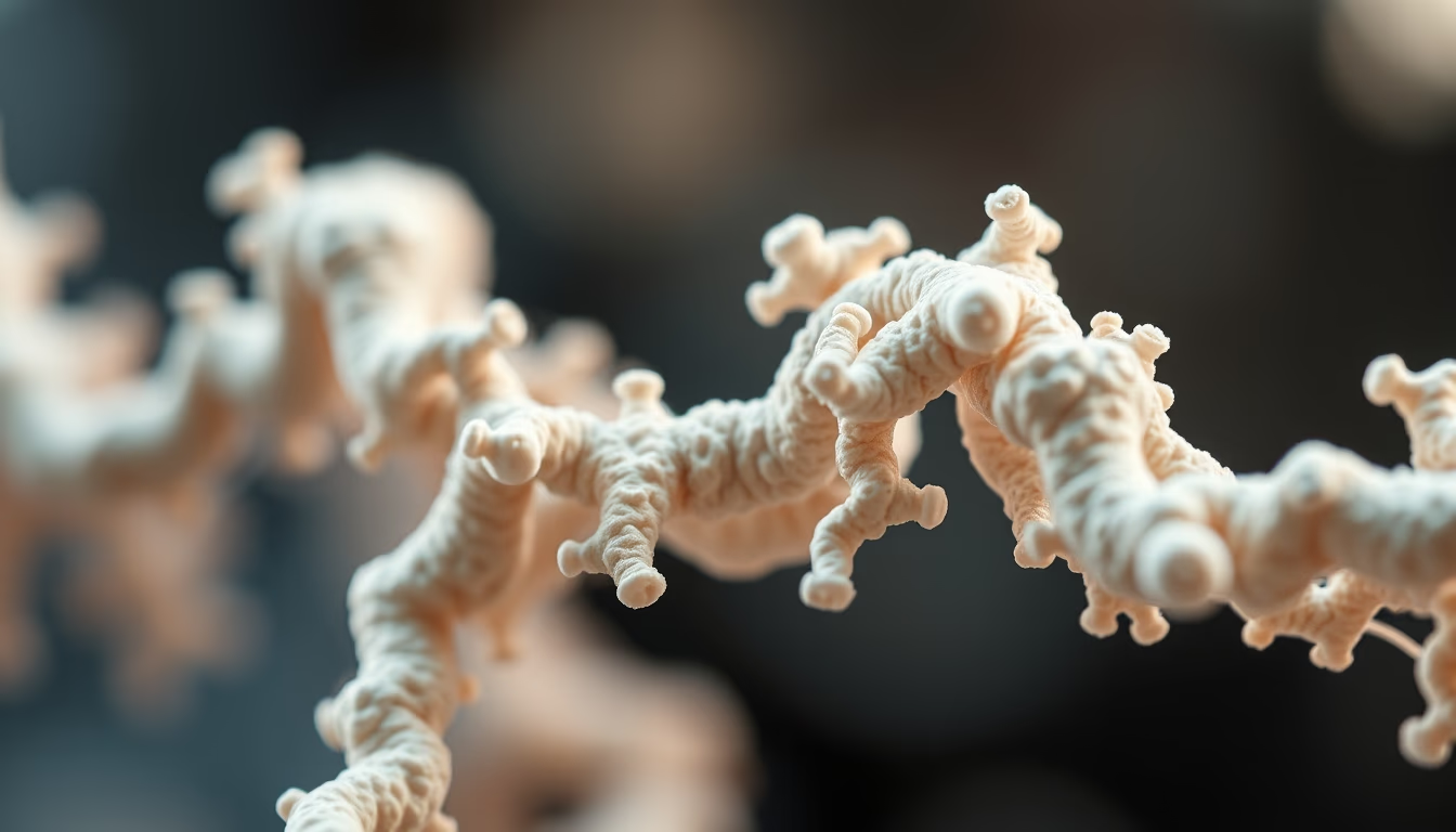
Proteins are essential macromolecules for life, whose function directly depends on their three-dimensional structure.
About this subject
Proteins are polymers made of amino acids, whose linear sequence is called the primary structure. This linear chain folds into alpha helices and beta sheets, forming the secondary structure. Interactions between side chains of amino acids far apart in the sequence generate the tertiary structure, a unique three-dimensional arrangement that determines the protein's function. Many functional proteins are composed of multiple subunits, configuring the quaternary structure.
Determining the three-dimensional structure of proteins is crucial for understanding their mechanisms of action and developing drugs. Techniques such as X-ray crystallography, cryo-electron microscopy, and nuclear magnetic resonance allow visualization of atoms and chemical groups in specific positions. For example, the structure of hemoglobin, discovered by Max Perutz, revealed how the protein transports oxygen and how mutations can cause diseases like sickle cell anemia.
Improper protein folding is associated with several neurodegenerative diseases, such as Alzheimer's, Parkinson's, and prion diseases. In these cases, proteins acquire abnormal conformations that aggregate, forming toxic plaques or fibrils. Understanding the normal and pathological three-dimensional structure is essential to design therapies that prevent or reverse these aggregates.
Computational protein modeling, such as AlphaFold, has revolutionized structure prediction from amino acid sequences. This accelerates research in structural biology and allows exploration of proteins from poorly studied organisms. The 3D structure is thus a bridge between genomics and biological function.
Frequently Asked Questions
What is the primary structure of a protein?
It is the linear sequence of amino acids that make up the protein, genetically determined. This sequence defines how the protein will fold into more complex structures.
How is the three-dimensional structure of a protein determined experimentally?
By methods such as X-ray crystallography, which analyzes diffraction patterns of protein crystals; cryo-electron microscopy, which reconstructs structures from images of frozen molecules; and nuclear magnetic resonance, which measures distances between atoms in solution.
Why is improper protein folding harmful?
Misfolded proteins can lose their normal function and form toxic aggregates, such as amyloid plaques. This is linked to neurodegenerative diseases like Alzheimer's and Parkinson's.
Direct URL
https://pub-c7d6a6ea828543ac903a74a341ccb2e1.r2.dev/imagens/protein-structure-3d-macro-closeup-p3.avifHow to credit
Include a visible link back to UtilizAí. Copy one of the snippets below:
<a href="https://xn--utiliza-eza.com/en/midia/imagens/protein-structure-3d-macro-closeup-p3">Protein structure 3d</a> by <a href="https://xn--utiliza-eza.com">UtilizAí</a>, licensed under <a href="https://creativecommons.org/licenses/by/4.0/">CC BY 4.0</a>.
[Protein structure 3d](https://xn--utiliza-eza.com/en/midia/imagens/protein-structure-3d-macro-closeup-p3) by [UtilizAí](https://xn--utiliza-eza.com), CC BY 4.0
License: CC-BY-4.0





