Protein structure 3d
1344×768 · AVIF · CC BY 4.0
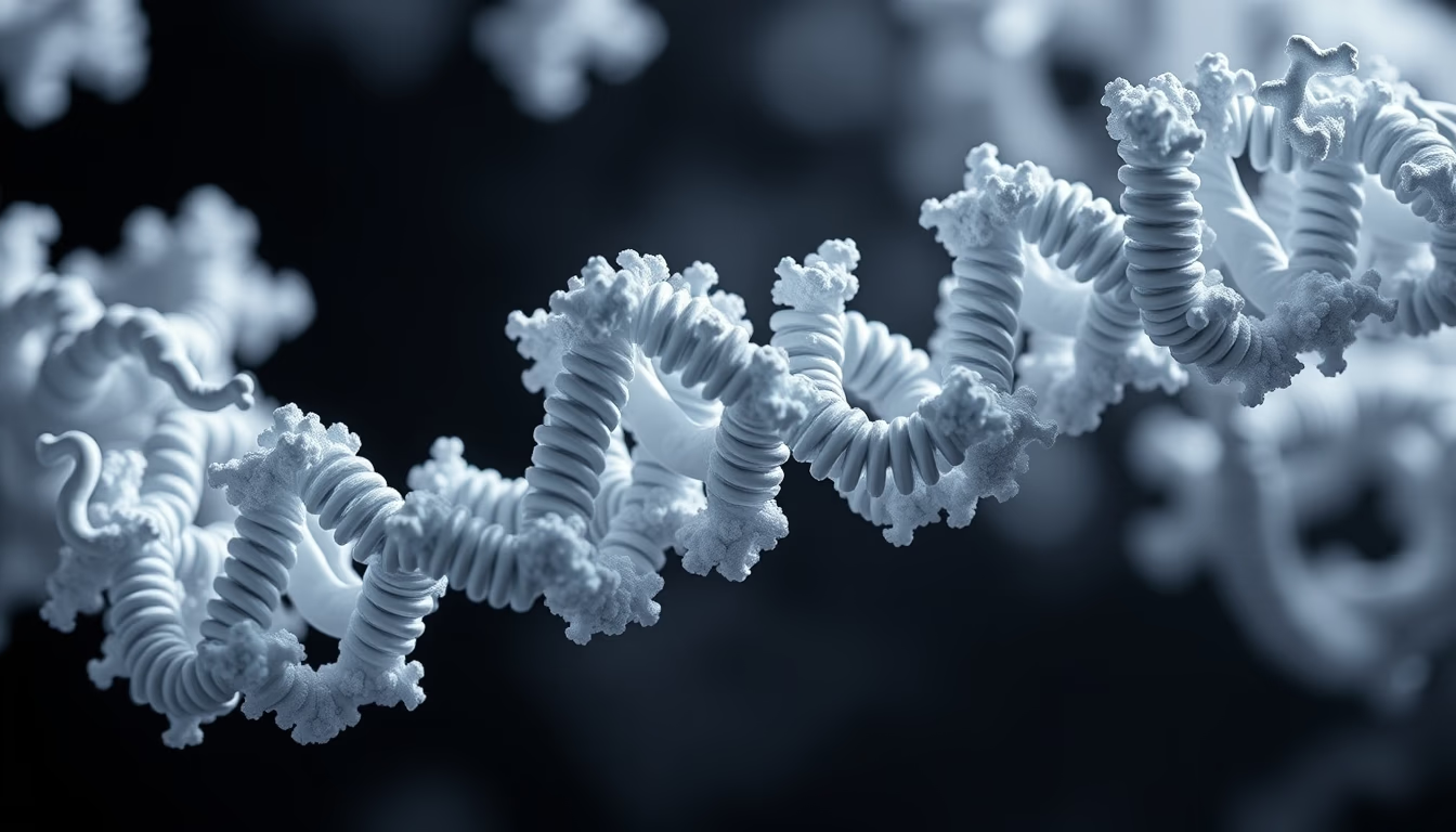
Three-dimensional protein structures reveal the molecular architecture that underpins life, essential for understanding diseases and developing drugs.
About this subject
Proteins are macromolecules responsible for nearly all cellular functions, from enzymatic catalysis to signaling and transport. Their function is intrinsically linked to their three-dimensional shape, determined by the amino acid sequence. The study of 3D protein structures began with X-ray crystallography, which in 1958 enabled the first determination of myoglobin structure by John Kendrew and hemoglobin by Max Perutz, earning them the Nobel Prize in Chemistry in 1962. Since then, methods such as nuclear magnetic resonance (NMR) and, more recently, cryo-electron microscopy (cryo-EM) have revolutionized the field, allowing visualization of proteins at near-atomic resolution without the need for crystallization.
The importance of protein structures extends beyond basic biology. In medicine, knowledge of the three-dimensional conformation of target proteins is crucial for rational drug design. For instance, the structure of HIV protease guided the development of inhibitors that transformed AIDS from a fatal disease into a chronic condition. More recently, cryo-EM was key to elucidating the SARS-CoV-2 spike protein in 2020, accelerating vaccine creation. Additionally, databases like the Protein Data Bank (PDB) store over 200,000 structures, accessible to researchers worldwide.
Interestingly, computational prediction of protein structures has advanced enormously with AlphaFold, an AI system from Google DeepMind that in 2021 predicted structures for millions of proteins with accuracy comparable to experiments. This opened doors for large-scale studies, such as identifying new functions and interactions. Still, protein dynamics, how they move and change shape, remains a challenge. Spectroscopic techniques and molecular dynamics simulations complement static images, revealing the molecular dance occurring inside cells.
Frequently Asked Questions
How are three-dimensional protein structures determined experimentally?
The main techniques are X-ray crystallography, nuclear magnetic resonance (NMR), and cryo-electron microscopy (cryo-EM). X-ray crystallography requires protein crystals and provides electron density maps. NMR studies proteins in solution, while cryo-EM allows visualization of frozen proteins in their native state without crystallization.
Why is the 3D structure of a protein important for drug development?
Knowing the three-dimensional structure helps identify active sites and cavities where small molecules can bind. Based on this information, drugs can be designed to fit precisely and modulate protein function, increasing efficacy and reducing side effects.
What is AlphaFold and how has it changed protein research?
AlphaFold is an artificial intelligence system developed by Google DeepMind that predicts the three-dimensional structure of proteins from their amino acid sequence. It achieved accuracy comparable to experimental methods, vastly accelerating research and enabling the prediction of millions of previously unknown protein structures.
Direct URL
https://pub-c7d6a6ea828543ac903a74a341ccb2e1.r2.dev/imagens/protein-structure-3d-fine-art-photography-p5.avifHow to credit
Include a visible link back to UtilizAí. Copy one of the snippets below:
<a href="https://xn--utiliza-eza.com/en/midia/imagens/protein-structure-3d-fine-art-photography-p5">Protein structure 3d</a> by <a href="https://xn--utiliza-eza.com">UtilizAí</a>, licensed under <a href="https://creativecommons.org/licenses/by/4.0/">CC BY 4.0</a>.
[Protein structure 3d](https://xn--utiliza-eza.com/en/midia/imagens/protein-structure-3d-fine-art-photography-p5) by [UtilizAí](https://xn--utiliza-eza.com), CC BY 4.0
License: CC-BY-4.0





