Protein structure 3d
1344×768 · AVIF · CC BY 4.0
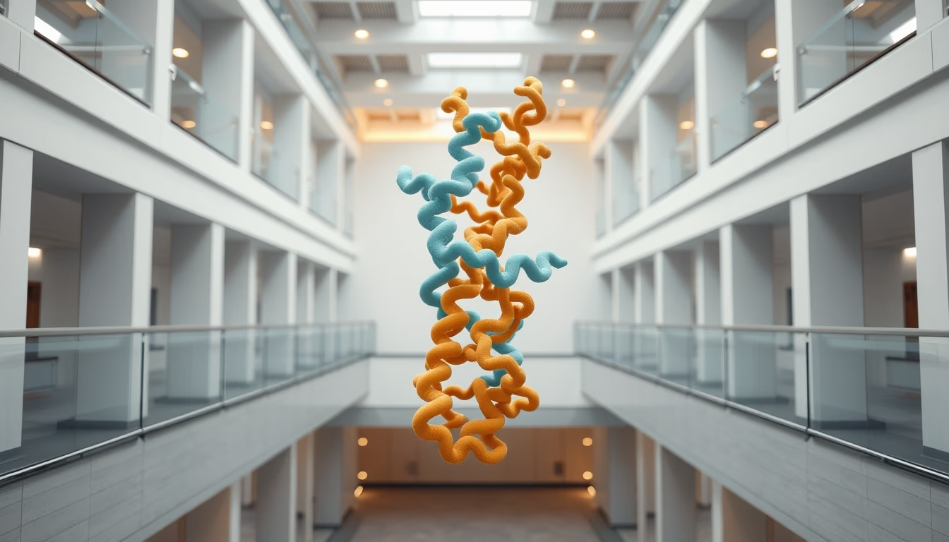
Three-dimensional protein structures reveal the molecular complexity that underpins life, combining data from X-ray crystallography and cryo-electron microscopy.
About this subject
Proteins are macromolecules essential for all living organisms, functioning as enzymes, antibodies, structural components, and transporters. Their biological function is directly linked to their three-dimensional structure, which is determined by the amino acid sequence (primary structure) and folding into alpha helices and beta sheets (secondary structure). The final spatial arrangement, called tertiary structure, is stabilized by interactions such as hydrogen bonds, hydrophobic interactions, and disulfide bridges. Many proteins further assemble into larger complexes, forming quaternary structure, such as hemoglobin, which carries oxygen in blood.
The study of protein three-dimensional structures has advanced tremendously with techniques like X-ray crystallography, which uses X-ray diffraction by protein crystals to generate electron density maps. More recently, cryo-electron microscopy (cryo-EM) has made it possible to visualize proteins in solution without crystallization, revolutionizing structural biology. In 2020, the Nobel Prize in Chemistry was awarded to Emmanuelle Charpentier and Jennifer Doudna for developing the CRISPR-Cas9 gene-editing technique, which relies on the structural understanding of the Cas9 protein.
Understanding protein structures is fundamental for rational drug design, protein engineering, and biotechnology. Databases like the Protein Data Bank (PDB) store thousands of three-dimensional models, accessible to researchers worldwide. Artificial intelligence, such as AlphaFold from Google DeepMind, can now predict protein structures with high accuracy, accelerating scientific discovery. Visualizations of these structures often use ribbon or surface representations that resemble architectural designs, hence the association with architectural photography in some scientific imagery.
Frequently Asked Questions
What determines the three-dimensional structure of a protein?
The three-dimensional structure is determined by the amino acid sequence (primary structure) and folding, either spontaneous or chaperone-assisted, guided by chemical interactions such as hydrogen bonds, hydrophobic effects, and disulfide bridges.
How are protein structures experimentally visualized?
The main techniques are X-ray crystallography, nuclear magnetic resonance (NMR), and cryo-electron microscopy (cryo-EM). Each has advantages and limitations, but cryo-EM has become prominent for large complexes.
Why is the Protein Data Bank (PDB) important?
The PDB is a public archive storing thousands of three-dimensional structures of proteins and nucleic acids, providing essential data for research in structural biology, drug discovery, and biotechnology.
Direct URL
https://pub-c7d6a6ea828543ac903a74a341ccb2e1.r2.dev/imagens/protein-structure-3d-architectural-photography-p11.avifHow to credit
Include a visible link back to UtilizAí. Copy one of the snippets below:
<a href="https://xn--utiliza-eza.com/en/midia/imagens/protein-structure-3d-architectural-photography-p11">Protein structure 3d</a> by <a href="https://xn--utiliza-eza.com">UtilizAí</a>, licensed under <a href="https://creativecommons.org/licenses/by/4.0/">CC BY 4.0</a>.
[Protein structure 3d](https://xn--utiliza-eza.com/en/midia/imagens/protein-structure-3d-architectural-photography-p11) by [UtilizAí](https://xn--utiliza-eza.com), CC BY 4.0
License: CC-BY-4.0





