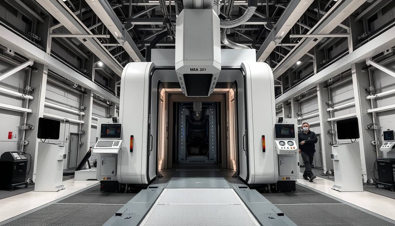
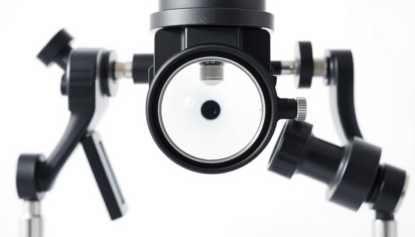
Orthopedic xray machine
1344×768 · AVIF · CC BY 4.0
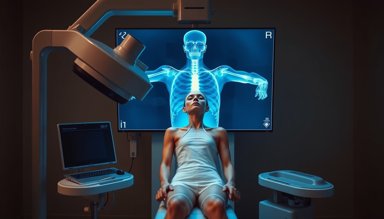
Orthopedic X-ray machines are essential for diagnosing fractures and bone diseases, combining imaging technology with medical precision.
About this subject
Orthopedic X-ray machines are fundamental tools in modern medicine, dedicated to visualizing the skeletal system. Unlike general radiography equipment, these devices are optimized to capture high-resolution images of bones and joints, aiding in the diagnosis of fractures, dislocations, arthritis, bone tumors, and congenital deformities. The technology relies on controlled emission of X-rays that pass through the body and are absorbed at different intensities by tissues, with bones being the densest and therefore most visible in resulting images.
The evolution of these machines has followed advances in radiology: from early analog devices using film plates to modern digital systems that allow instant image acquisition and processing. Currently, equipment such as the surgical C-arm is widely used in minimally invasive orthopedic procedures, providing real-time fluoroscopy to guide implant placement or fracture reduction. Radiation dose is strictly controlled to ensure patient and staff safety, following protocols like ALARA (As Low As Reasonably Achievable).
In Brazil, the use of orthopedic X-rays is standardized by the Ministry of Health and the National Health Surveillance Agency (Anvisa), which establish technical and radiological protection requirements. Public and private hospitals rely on these machines in emergency rooms and surgical centers. A curious fact is that the first orthopedic radiograph in history was performed by Wilhelm Röntgen in 1895, of his wife's hand, paving the way for modern orthopedics. Despite advances in other techniques such as computed tomography and magnetic resonance imaging, the orthopedic X-ray remains the first-line exam due to its low cost, speed, and accessibility.
Frequently Asked Questions
What is the difference between an orthopedic X-ray and a regular X-ray?
An orthopedic X-ray is specifically designed for bones and joints, with dose and positioning adjustments that prioritize detailed visualization of the skeleton. A regular X-ray can be used for chest, abdomen, or other areas with different parameters.
Is it safe to have frequent orthopedic X-ray exams?
Yes, as long as established dose limits are respected. Exposure in an orthopedic X-ray is low (equivalent to a few days of natural background radiation). Doctors assess the real need for each exam to avoid unnecessary repetitions.
How does an orthopedic X-ray machine work?
It emits a beam of X-rays that passes through the body. Bones absorb more radiation and appear white on the image (radiolucent). Digital sensors or film capture the remaining radiation, producing a radiograph that aids diagnosis.
Direct URL
https://pub-c7d6a6ea828543ac903a74a341ccb2e1.r2.dev/imagens/orthopedic-xray-machine-high-fashion-editorial-p6.avifHow to credit
Include a visible link back to UtilizAí. Copy one of the snippets below:
<a href="https://xn--utiliza-eza.com/en/midia/imagens/orthopedic-xray-machine-high-fashion-editorial-p6">Orthopedic xray machine</a> by <a href="https://xn--utiliza-eza.com">UtilizAí</a>, licensed under <a href="https://creativecommons.org/licenses/by/4.0/">CC BY 4.0</a>.
[Orthopedic xray machine](https://xn--utiliza-eza.com/en/midia/imagens/orthopedic-xray-machine-high-fashion-editorial-p6) by [UtilizAí](https://xn--utiliza-eza.com), CC BY 4.0
License: CC-BY-4.0