Nucleus chromosomes microscope
1344×768 · AVIF · CC BY 4.0
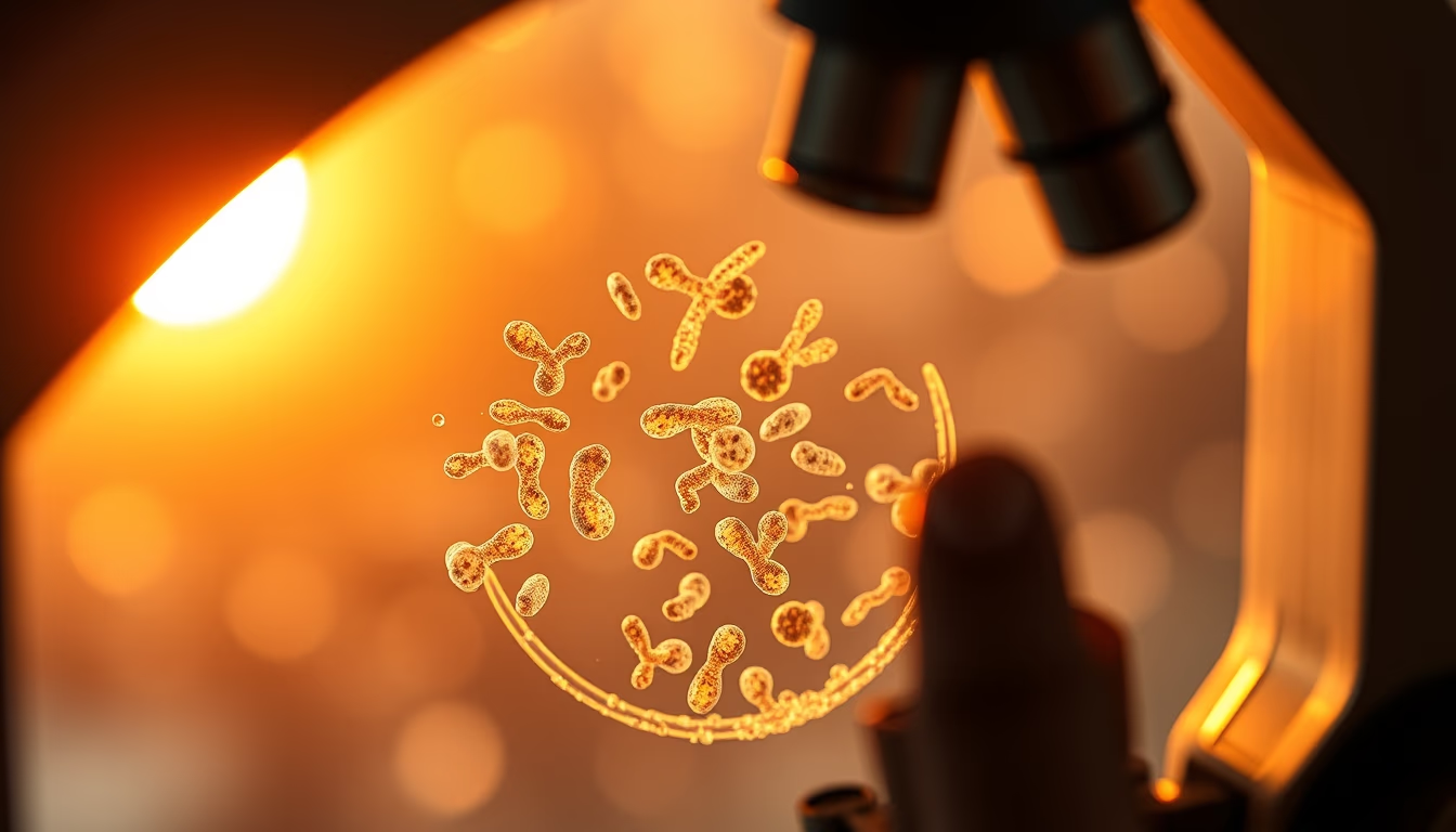
Cell nucleus and chromosomes under the microscope: structure, function, and importance for genetics.
About this subject
The cell nucleus is the organelle that houses the cell's genetic material, organized into chromosomes. During interphase, chromosomes are decondensed as chromatin, but during cell division they compact into structures visible under a light microscope. Fluorescence, confocal, and electron microscopy allow studying nuclear morphology and chromosome dynamics in real time.
Each species has a characteristic number of chromosomes. For example, humans have 46 chromosomes (23 pairs). Chromosomal abnormalities, such as trisomies or deletions, are associated with genetic diseases like Down syndrome and cancer. Cytogenetics uses banding techniques to identify each chromosome individually.
The nucleus also has a nuclear envelope with pores that regulate molecule traffic between nucleus and cytoplasm. The nucleolus, a nuclear substructure, is responsible for ribosomal RNA synthesis. Atomic force microscopy and cryo-electron microscopy reveal molecular details of chromatin and the nuclear envelope.
Studying the nucleus and chromosomes is fundamental for understanding heredity, gene regulation, and pathological processes. Techniques such as FISH (fluorescence in situ hybridization) allow localizing specific genes on chromosomes. Microscopy images are essential for diagnostics and research in cell and molecular biology.
Frequently Asked Questions
How many chromosomes do humans have?
Humans have 46 chromosomes, organized into 23 pairs. Each pair is inherited one from the mother and one from the father.
What is the difference between chromatin and chromosomes?
Chromatin is the uncondensed form of DNA during interphase, while chromosomes are the highly condensed form visible during cell division.
What type of microscope is used to see chromosomes?
Chromosomes can be seen with a light microscope after staining. Fine details require electron or fluorescence microscopy.
Direct URL
https://pub-c7d6a6ea828543ac903a74a341ccb2e1.r2.dev/imagens/nucleus-chromosomes-microscope-travel-photography-p6.avifHow to credit
Include a visible link back to UtilizAí. Copy one of the snippets below:
<a href="https://xn--utiliza-eza.com/en/midia/imagens/nucleus-chromosomes-microscope-travel-photography-p6">Nucleus chromosomes microscope</a> by <a href="https://xn--utiliza-eza.com">UtilizAí</a>, licensed under <a href="https://creativecommons.org/licenses/by/4.0/">CC BY 4.0</a>.
[Nucleus chromosomes microscope](https://xn--utiliza-eza.com/en/midia/imagens/nucleus-chromosomes-microscope-travel-photography-p6) by [UtilizAí](https://xn--utiliza-eza.com), CC BY 4.0
License: CC-BY-4.0





