Nucleus chromosomes microscope
1344×768 · AVIF · CC BY 4.0
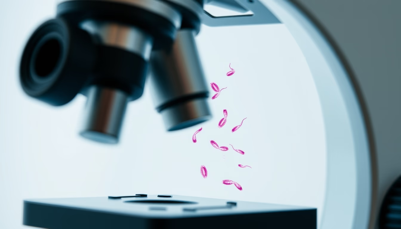
Chromosomes in the cell nucleus observed under a microscope, revealing the organization of genetic material during cell division.
About this subject
Chromosomes are thread-like structures composed of DNA and proteins, found in the nucleus of eukaryotic cells. They carry genes that determine hereditary traits and control cellular functions. During most of the cell cycle, DNA is uncoiled as chromatin, but during mitosis and meiosis it condenses into chromosomes visible under a light microscope. This condensation allows accurate segregation of genetic material into daughter cells.
Microscopic observation of chromosomes became possible through staining techniques, such as Giemsa staining, which produces characteristic banding patterns on each chromosome. These patterns are used in karyotypes to identify chromosomal abnormalities, such as Down, Turner, and Klinefelter syndromes. Cytogenetics, the branch of genetics that studies chromosomes, began in the late 19th century with Walther Flemming's observations of mitosis in salamander cells.
In humans, the diploid number of chromosomes is 46, organized into 23 pairs. Each pair consists of one chromosome inherited from the father and one from the mother. Modern microscopy, including confocal and electron microscopy, reveals details of chromosomal structure such as the centromere and telomeres. The lo-fi disposable camera effect in the image adds a nostalgic aesthetic that contrasts with the technical precision of science, yet still evokes the beauty of cell biology.
Frequently Asked Questions
What are chromosomes?
Chromosomes are structures in the cell nucleus made of DNA and proteins that carry genes. They condense during cell division to ensure accurate distribution of genetic material.
How are chromosomes visualized under a microscope?
Chromosomes are stained with specific dyes, such as Giemsa, and observed under light microscopes during metaphase of mitosis, when they are most condensed.
How many chromosomes are in the human nucleus?
Normal human cells have 46 chromosomes, organized into 23 pairs, one from each parent.
Direct URL
https://pub-c7d6a6ea828543ac903a74a341ccb2e1.r2.dev/imagens/nucleus-chromosomes-microscope-lo-fi-disposable-camera-p11.avifHow to credit
Include a visible link back to UtilizAí. Copy one of the snippets below:
<a href="https://xn--utiliza-eza.com/en/midia/imagens/nucleus-chromosomes-microscope-lo-fi-disposable-camera-p11">Nucleus chromosomes microscope</a> by <a href="https://xn--utiliza-eza.com">UtilizAí</a>, licensed under <a href="https://creativecommons.org/licenses/by/4.0/">CC BY 4.0</a>.
[Nucleus chromosomes microscope](https://xn--utiliza-eza.com/en/midia/imagens/nucleus-chromosomes-microscope-lo-fi-disposable-camera-p11) by [UtilizAí](https://xn--utiliza-eza.com), CC BY 4.0
License: CC-BY-4.0





