Nucleus chromosomes microscope
1344×768 · AVIF · CC BY 4.0
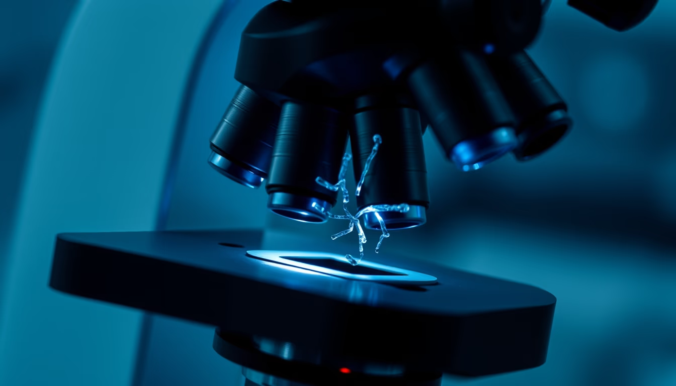
The cell nucleus houses chromosomes, DNA structures carrying genetic information. Advanced microscopy reveals their complex organization.
About this subject
The cell nucleus is a membrane-bound organelle in eukaryotic cells that stores and protects genetic material. Inside, chromosomes organize as highly compacted DNA strands wrapped around histone proteins, allowing meters of DNA to fit into a microscopic space. Chromosome structure changes during the cell cycle: during interphase they are less condensed as chromatin; during cell division they condense into forms visible under light microscopy.
Fluorescence microscopy and cryo-electron microscopy are essential for visualizing chromosomes and the nucleus. Specific dyes like DAPI bind to DNA and highlight chromosomes under ultraviolet light. Confocal microscopy enables three-dimensional reconstructions of the nucleus, revealing chromosome territories and gene positioning. Such images are crucial for genetics research, cancer studies, and hereditary diseases.
Interestingly, the number and shape of chromosomes are species-specific. Humans have 46 chromosomes in pairs, while some ants have only 2. Chromosomal abnormalities like trisomies or deletions are linked to genetic syndromes. Chromosome analysis via microscopy is a cornerstone of cytogenetic diagnosis.
The intersection of science and art, as hinted by the high-fashion editorial style, can highlight the intrinsic beauty of cellular structures. Nucleus and chromosome microscope images with vivid staining or digital processing transcend the lab to become visual works communicating life's complexity.
Frequently Asked Questions
What is the cell nucleus and what is its function?
The cell nucleus is an organelle containing the cell's DNA, controlling activities like growth, metabolism, and reproduction. It is enclosed by a nuclear membrane with pores regulating material exchange.
How are chromosomes visualized under a microscope?
Chromosomes are stained with specific dyes like Giemsa or DAPI that bind to DNA. During metaphase they are most condensed and visible under light microscopy. Techniques like FISH use fluorescent probes to label specific sequences.
How many chromosomes do humans have?
Humans have 46 chromosomes arranged in 23 pairs. One pair are sex chromosomes (XX or XY). Changes in number, such as Down syndrome (trisomy 21), cause genetic conditions.
Direct URL
https://pub-c7d6a6ea828543ac903a74a341ccb2e1.r2.dev/imagens/nucleus-chromosomes-microscope-high-fashion-editorial-p8.avifHow to credit
Include a visible link back to UtilizAí. Copy one of the snippets below:
<a href="https://xn--utiliza-eza.com/en/midia/imagens/nucleus-chromosomes-microscope-high-fashion-editorial-p8">Nucleus chromosomes microscope</a> by <a href="https://xn--utiliza-eza.com">UtilizAí</a>, licensed under <a href="https://creativecommons.org/licenses/by/4.0/">CC BY 4.0</a>.
[Nucleus chromosomes microscope](https://xn--utiliza-eza.com/en/midia/imagens/nucleus-chromosomes-microscope-high-fashion-editorial-p8) by [UtilizAí](https://xn--utiliza-eza.com), CC BY 4.0
License: CC-BY-4.0





