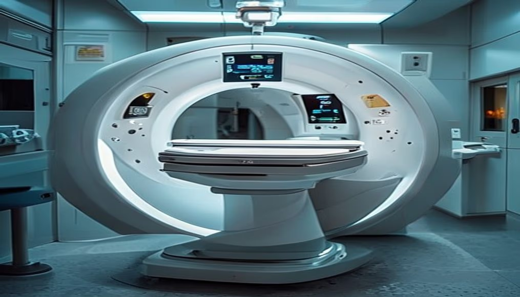
Mri scanner machine in radiology department
1344×768 · AVIF · CC BY 4.0
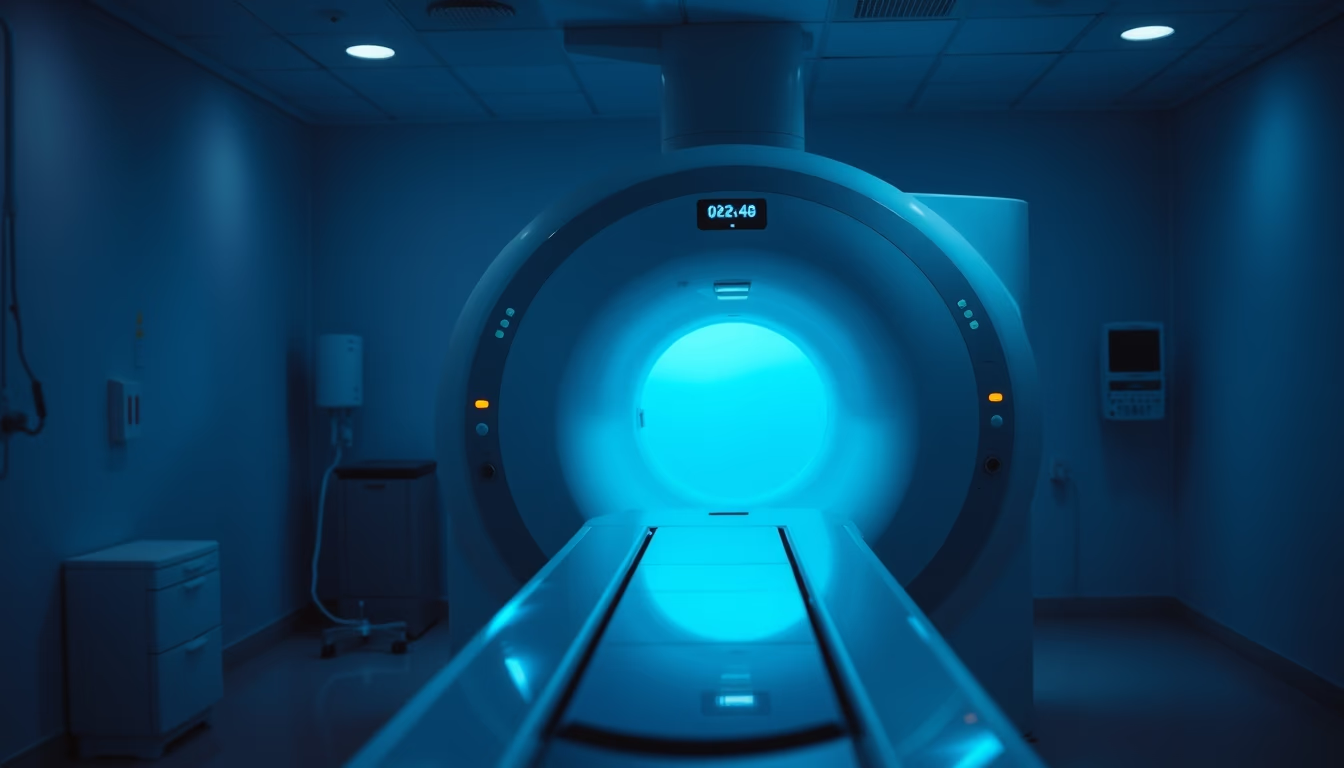
Magnetic resonance imaging is a noninvasive exam that uses magnetic fields and radio waves to produce detailed images of internal organs and tissues.
About this subject
The magnetic resonance imaging (MRI) machine is one of the most advanced tools in modern radiology. Unlike computed tomography, which uses ionizing radiation, MRI employs a strong magnetic field (typically 1.5 to 3 Tesla) and radiofrequency pulses to align hydrogen protons in the body and then capture signals emitted as these protons return to their original state. This physical principle, known as nuclear magnetic resonance, was discovered in the 1940s and applied to medicine from the 1970s, with the first clinical scanners appearing in the early 1980s.
The radiology department housing the machine follows strict safety protocols. Due to the intense magnetic field, ferromagnetic metallic objects (such as keys, credit cards, and electronic devices) are prohibited in the exam room. Patients with metal implants, such as pacemakers or aneurysm clips, may be contraindicated for the procedure. The exam is particularly useful for visualizing the central nervous system, joints, abdomen, and pelvis, offering exceptional soft tissue contrast. In Brazil, MRI is widely available in large hospitals and specialized clinics, being one of the most requested tests for diagnosing tumors, neurological lesions, and musculoskeletal diseases.
A technical curiosity: the loud, characteristic noise during the exam (reaching 120 decibels) is caused by vibration of the gradient coils due to rapid electrical current variations. Therefore, patients receive earplugs or headphones. Additionally, functional MRI (fMRI) allows mapping of brain activity in real time, being an essential tool in neuroscience. The image, captured during the twilight blue hour, shows the equipment in a low-light environment, highlighting its symmetrical design and the serene atmosphere of the diagnostic imaging sector.
Frequently Asked Questions
What is the difference between MRI and CT scan?
MRI uses magnetic fields and radio waves without ionizing radiation, ideal for soft tissues. CT uses X-rays, providing better visualization of bones and calcified structures.
How long does an MRI exam take?
The exam can last from 15 to 60 minutes, depending on the body region and need for contrast. The patient must remain still to avoid image artifacts.
Is MRI safe for pregnant women?
Generally, MRI is not recommended in the first trimester unless essential. From the second trimester onward, it can be performed safely, but always assessing risks and benefits.
Direct URL
https://pub-c7d6a6ea828543ac903a74a341ccb2e1.r2.dev/imagens/mri-scanner-machine-in-radiology-department-symmetric-composition-twilight-blue-hour.avifHow to credit
Include a visible link back to UtilizAí. Copy one of the snippets below:
<a href="https://xn--utiliza-eza.com/en/midia/imagens/mri-scanner-machine-in-radiology-department-symmetric-composition-twilight-blue-hour">Mri scanner machine in radiology department</a> by <a href="https://xn--utiliza-eza.com">UtilizAí</a>, licensed under <a href="https://creativecommons.org/licenses/by/4.0/">CC BY 4.0</a>.
[Mri scanner machine in radiology department](https://xn--utiliza-eza.com/en/midia/imagens/mri-scanner-machine-in-radiology-department-symmetric-composition-twilight-blue-hour) by [UtilizAí](https://xn--utiliza-eza.com), CC BY 4.0
License: CC-BY-4.0